Podcast
Questions and Answers
What is the primary function of the psoas minor muscle?
What is the primary function of the psoas minor muscle?
- Strong flexion of the trunk
- Extension of the lumbar spine
- Weak flexion of the trunk (correct)
- Rotation of the pelvis
At which vertebral level does the abdominal aorta bifurcate into the common iliac arteries?
At which vertebral level does the abdominal aorta bifurcate into the common iliac arteries?
- L2
- L1
- L3
- L4 (correct)
Which branch of the abdominal aorta arises at the level of the L2 vertebrae?
Which branch of the abdominal aorta arises at the level of the L2 vertebrae?
- Celiac trunk
- Superior mesenteric artery
- Renal artery
- Inferior mesenteric artery (correct)
Which artery supplies the second half of the large intestine?
Which artery supplies the second half of the large intestine?
Which of the following arteries is not a branch of the celiac trunk?
Which of the following arteries is not a branch of the celiac trunk?
Which nerve root innervates the psoas minor muscle?
Which nerve root innervates the psoas minor muscle?
Which artery supplies the kidneys?
Which artery supplies the kidneys?
What is the level of the vertebral column where the abdominal aorta enters the abdominal cavity?
What is the level of the vertebral column where the abdominal aorta enters the abdominal cavity?
What is the origin of the internal oblique muscle?
What is the origin of the internal oblique muscle?
What is the action of the transversus abdominis muscle?
What is the action of the transversus abdominis muscle?
What is the purpose of the conjoint tendon?
What is the purpose of the conjoint tendon?
What is the insertion of the rectus abdominis muscle?
What is the insertion of the rectus abdominis muscle?
What is the innervation of the internal oblique muscle?
What is the innervation of the internal oblique muscle?
What is the characteristic of the posterior border of the external oblique muscle?
What is the characteristic of the posterior border of the external oblique muscle?
What is the significance of the linea semilunaris?
What is the significance of the linea semilunaris?
What is the purpose of the tendinous intersections of the rectus abdominis muscle?
What is the purpose of the tendinous intersections of the rectus abdominis muscle?
What type of muscle is the dartos muscle?
What type of muscle is the dartos muscle?
What is the main function of the external oblique muscle?
What is the main function of the external oblique muscle?
What is the insertion point of the external oblique muscle?
What is the insertion point of the external oblique muscle?
Which nerve innervates the external oblique muscle?
Which nerve innervates the external oblique muscle?
What is the deepest layer of fascia in the scrotum?
What is the deepest layer of fascia in the scrotum?
What is the function of the deep fascia?
What is the function of the deep fascia?
Which muscle is located on either side of the midline anteriorly?
Which muscle is located on either side of the midline anteriorly?
What is the function of Colles' fascia?
What is the function of Colles' fascia?
What forms the posterior wall of the rectus sheath above the costal margin?
What forms the posterior wall of the rectus sheath above the costal margin?
Which of the following muscles forms the anterior wall of the rectus sheath between the level of the arcuate line and the pubis?
Which of the following muscles forms the anterior wall of the rectus sheath between the level of the arcuate line and the pubis?
What is the purpose of an aponeurosis?
What is the purpose of an aponeurosis?
What is the function of the linea alba?
What is the function of the linea alba?
What is the transversalis fascia?
What is the transversalis fascia?
What is the level of the arcuate line in relation to the anterior superior iliac spine?
What is the level of the arcuate line in relation to the anterior superior iliac spine?
What is the function of the aponeurosis of the external oblique above the costal margin?
What is the function of the aponeurosis of the external oblique above the costal margin?
What is the relationship between an aponeurosis and a tendon?
What is the relationship between an aponeurosis and a tendon?
Where do the gonadal arteries originate from?
Where do the gonadal arteries originate from?
What is the purpose of the lumbar arteries?
What is the purpose of the lumbar arteries?
What are the final branches of the abdominal aorta?
What are the final branches of the abdominal aorta?
Where does the inferior vena cava form?
Where does the inferior vena cava form?
What structures do the common iliac arteries supply?
What structures do the common iliac arteries supply?
What drains the structures of the posterior abdominal wall?
What drains the structures of the posterior abdominal wall?
Where does the inferior vena cava enter the heart?
Where does the inferior vena cava enter the heart?
What is the destination of the blood from the gut tube?
What is the destination of the blood from the gut tube?
Flashcards are hidden until you start studying
Study Notes
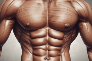
Muscles of the Anterior Abdominal Wall
- The muscles of the anterior abdominal wall consist of three broad, thin sheets of muscle that are most pronounced laterally and become aponeurotic anteriorly.
- The four main muscles are: external oblique, internal oblique, transversus abdominis, and rectus abdominis.
External Oblique
- Origin: lower eight ribs
- Insertion: Xiphoid process, linea alba, iliac crest, pubic crest, and tubercle
- Action: support and compress abdominal contents, assist in flexion and rotation of trunk, assist in forced expiration, micturition, defecation, and vomiting
- Innervation: lower six thoracic nerves, iliohypogastric and ilioinguinal nerves (L1)
Internal Oblique
- Origin: iliac crest and lateral 2/3 of inguinal ligament
- Insertion: lower three ribs, Xiphoid process, linea alba, and symphysis pubis
- Action: same as external oblique
- Innervation: same as external oblique
Transversus Abdominis
- Origin: lower six costal cartilages, iliac crest, and lateral 2/3 of inguinal ligaments
- Insertion: Xiphoid process, linea alba, and symphysis pubis
- Action: compress abdominal contents
- Innervation: same as external oblique
Conjoint Tendon
- Formed by the lowest tendinous fibers (aponeurosis) of internal oblique and transversus abdominis muscles
- Attaches medially to the linea alba, but has a lateral free border
- Has an essential role in protecting a weak area in the abdominal wall, where a weakening of the conjoint tendon may lead to a direct inguinal hernia
Rectus Abdominis
- Origin: symphysis pubis and pubic crest
- Insertion: 5th, 6th, and 7th costal cartilages and xiphoid process
- Action: compress, flex vertebral column, and accessory muscle of expiration
- Innervation: lower six thoracic nerves
Rectus Sheath
- The composition of the walls of the rectus sheath changes at different levels
- The rectus sheath is generally considered at three levels: above the costal margin, between the costal margin and the arcuate line, and between the level of the arcuate line and the pubis
- The aponeuroses of the external oblique, internal oblique, and transversus abdominis muscles form the rectus sheath
Linea Alba
- A fibrous band that separates the two rectus sheaths from each other
- Extends from the xiphoid process to the symphysis pubis
- Formed by the fusion of the aponeuroses of the lateral muscles of the two sides
Abdominal Aorta
- Enters the abdomen through the aortic aperture of the diaphragm (T12)
- Branches to the diaphragm (inferior phrenic), abdominal wall (lumbar, median sacral), abdominal viscera, kidneys, and ovaries/testes
- Bifurcates at L4 into common iliac arteries to supply the lower limb and pelvis
Arterial Branches of Abdominal Aorta
- Celiac trunk: the first branch of the abdominal aorta, supplying the stomach, spleen, and liver
- Superior mesenteric artery: supplying most of the small intestine and the first half of the large intestine, or colon
- Inferior mesenteric artery: a small, unpaired artery supplying the second half of the large intestine
- Renal arteries: serving the kidneys
- Gonadal arteries: serving the ovaries and testes
- Lumbar arteries: serving the heavy muscles of the abdomen and trunk walls
Inferior Vena Cava
- Forms at L5 vertebral level when the left and right common iliac veins unite
- Drains the structures of the posterior abdominal wall, the kidneys, and the suprarenal glands
- The veins that drain blood from the gut tube pass their blood into the portal system, not the IVC
- Blood from the portal system passes through the liver before entering the IVC via hepatic veins
- The IVC passes from the liver and through the diaphragm at T8 before entering the inferior surface of the right atrium
Studying That Suits You
Use AI to generate personalized quizzes and flashcards to suit your learning preferences.


