Podcast
Questions and Answers
Which layer of the heart protects and anchors the heart, prevents overfilling with blood, and allows for relatively friction-free movement?
Which layer of the heart protects and anchors the heart, prevents overfilling with blood, and allows for relatively friction-free movement?
- Endocardium
- Epicardium
- Pericardium (correct)
- Myocardium
Which major vessels return blood to the heart?
Which major vessels return blood to the heart?
- Right and left pulmonary arteries
- Pulmonary trunk and ascending aorta
- Brachiocephalic, left common carotid, and subclavian arteries
- Superior and inferior venae cavae (correct)
Which arteries supply the heart with blood?
Which arteries supply the heart with blood?
- Right and left coronary arteries (correct)
- Right and left pulmonary arteries
- Subclavian and common carotid arteries
- Ascending aorta and brachiocephalic artery
Where is the heart located?
Where is the heart located?
What is the size of the heart approximately compared to?
What is the size of the heart approximately compared to?
Which layer forms the bulk of the heart and is composed of cardiac muscle?
Which layer forms the bulk of the heart and is composed of cardiac muscle?
Which vessels convey blood away from the heart?
Which vessels convey blood away from the heart?
What are the components of the conduction system of the heart?
What are the components of the conduction system of the heart?
Which vessels supply/drain the heart?
Which vessels supply/drain the heart?
What is the function of the fibrous skeleton of the heart?
What is the function of the fibrous skeleton of the heart?
Which veins return blood to the heart in the posterior view?
Which veins return blood to the heart in the posterior view?
Which arteries supply the heart in the posterior view?
Which arteries supply the heart in the posterior view?
What are the layers that make up the heart wall?
What are the layers that make up the heart wall?
What marks the walls of the atria?
What marks the walls of the atria?
What is the functional blood supply to the heart muscle itself called?
What is the functional blood supply to the heart muscle itself called?
What makes the left side of the heart have a thicker wall?
What makes the left side of the heart have a thicker wall?
What ensures unidirectional blood flow through the heart?
What ensures unidirectional blood flow through the heart?
What are the components of the heart's conduction system?
What are the components of the heart's conduction system?
What is the size of the heart approximately compared to?
What is the size of the heart approximately compared to?
What is the role of the atria in the heart?
What is the role of the atria in the heart?
What is the role of the ventricles in the heart?
What is the role of the ventricles in the heart?
What marks the walls of the ventricles?
What marks the walls of the ventricles?
What is the pathway of blood through the heart and lungs primarily involves?
What is the pathway of blood through the heart and lungs primarily involves?
What type of muscle is the cardiac muscle?
What type of muscle is the cardiac muscle?
What is the approximate location of the heart?
What is the approximate location of the heart?
Which vessels supply/drain the heart in the anterior view?
Which vessels supply/drain the heart in the anterior view?
What is the function of the fibrous skeleton of the heart?
What is the function of the fibrous skeleton of the heart?
Which vessels return blood to the heart in the posterior view?
Which vessels return blood to the heart in the posterior view?
What are the coverings of the heart?
What are the coverings of the heart?
What is the role of the papillary muscles in the heart?
What is the role of the papillary muscles in the heart?
Which blood vessels ensure blood delivery to the heart muscle even if major vessels are occluded?
Which blood vessels ensure blood delivery to the heart muscle even if major vessels are occluded?
What is the function of intercalated discs in cardiac muscle?
What is the function of intercalated discs in cardiac muscle?
What is the primary function of the atrioventricular (AV) and semilunar valves in the heart?
What is the primary function of the atrioventricular (AV) and semilunar valves in the heart?
What is the role of the SA node in the heart's conduction system?
What is the role of the SA node in the heart's conduction system?
True or false: The fibrous skeleton of the heart is primarily composed of cardiac muscle.
True or false: The fibrous skeleton of the heart is primarily composed of cardiac muscle.
True or false: The ascending aorta splits into the right and left pulmonary arteries.
True or false: The ascending aorta splits into the right and left pulmonary arteries.
True or false: The pericardium allows for the heart to work in a relatively friction-free environment.
True or false: The pericardium allows for the heart to work in a relatively friction-free environment.
True or false: The left and right pulmonary veins convey blood away from the heart.
True or false: The left and right pulmonary veins convey blood away from the heart.
True or false: The external heart in the anterior view shows the great cardiac vein and the posterior vein to the left ventricle.
True or false: The external heart in the anterior view shows the great cardiac vein and the posterior vein to the left ventricle.
Coronary circulation is the only blood supply to the heart muscle itself
Coronary circulation is the only blood supply to the heart muscle itself
The atria have papillary muscles marking their walls
The atria have papillary muscles marking their walls
The left side of the heart has a thinner wall due to the lower pressure needed to pump blood to the entire body
The left side of the heart has a thinner wall due to the lower pressure needed to pump blood to the entire body
The epicardium is the innermost layer of the heart wall
The epicardium is the innermost layer of the heart wall
The SA node is responsible for initiating the heartbeat
The SA node is responsible for initiating the heartbeat
Flashcards are hidden until you start studying
Study Notes
Anatomy and Physiology of the Heart
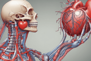
- The heart is located in the thoracic cavity, between the lungs and behind the sternum
- The chambers of the heart include the atria (receiving chambers) and the ventricles (discharging chambers)
- The blood vessels that connect to the heart are the superior and inferior venae cavae, pulmonary arteries, pulmonary veins, and the aorta
- The layers that make up the heart wall are the epicardium, myocardium, and endocardium
- The left side of the heart has a thicker wall due to the higher pressure needed to pump blood to the entire body
- The atria have protruding auricles and pectinate muscles mark atrial walls
- The ventricles have papillary muscles and trabeculae carneae muscles marking their walls
- The pathway of blood through the heart and lungs involves the atria, ventricles, valves, and major blood vessels
- Coronary circulation is the functional blood supply to the heart muscle itself, with collateral routes ensuring blood delivery even if major vessels are occluded
- Heart valves, including atrioventricular and semilunar valves, ensure unidirectional blood flow through the heart
- Cardiac muscle is striated, short, fat, branched, and interconnected, with intercalated discs anchoring cardiac cells together and allowing free passage of ions
- The heart's conduction system involves the SA node, AV node, atrioventricular bundle, bundle branches, and Purkinje fibers, and it is related to electrocardiography and the cardiac cycle
Anatomy and Physiology of the Heart
- The heart is located in the thoracic cavity, between the lungs and behind the sternum
- The chambers of the heart include the atria (receiving chambers) and the ventricles (discharging chambers)
- The blood vessels that connect to the heart are the superior and inferior venae cavae, pulmonary arteries, pulmonary veins, and the aorta
- The layers that make up the heart wall are the epicardium, myocardium, and endocardium
- The left side of the heart has a thicker wall due to the higher pressure needed to pump blood to the entire body
- The atria have protruding auricles and pectinate muscles mark atrial walls
- The ventricles have papillary muscles and trabeculae carneae muscles marking their walls
- The pathway of blood through the heart and lungs involves the atria, ventricles, valves, and major blood vessels
- Coronary circulation is the functional blood supply to the heart muscle itself, with collateral routes ensuring blood delivery even if major vessels are occluded
- Heart valves, including atrioventricular and semilunar valves, ensure unidirectional blood flow through the heart
- Cardiac muscle is striated, short, fat, branched, and interconnected, with intercalated discs anchoring cardiac cells together and allowing free passage of ions
- The heart's conduction system involves the SA node, AV node, atrioventricular bundle, bundle branches, and Purkinje fibers, and it is related to electrocardiography and the cardiac cycle
Studying That Suits You
Use AI to generate personalized quizzes and flashcards to suit your learning preferences.