Podcast
Questions and Answers
How many cranial bones are there in the skull?
How many cranial bones are there in the skull?
- 8 (correct)
- 6
- 14
- 22
What are the main sutures of the skull?
What are the main sutures of the skull?
Coronal, sagittal, lambdoid, squamosal
The metopic suture is always present in adults.
The metopic suture is always present in adults.
False (B)
What does the dura mater protect?
What does the dura mater protect?
What are the layers of the meninges from outermost to innermost?
What are the layers of the meninges from outermost to innermost?
What is the function of the meninges?
What is the function of the meninges?
The skull contains air ______ which are highly variable in appearance.
The skull contains air ______ which are highly variable in appearance.
The brain is located outside the cranial vault.
The brain is located outside the cranial vault.
What is hydrocephalus?
What is hydrocephalus?
Name one type of intracranial hemorrhage.
Name one type of intracranial hemorrhage.
Match the following structures with their functions:
Match the following structures with their functions:
Flashcards are hidden until you start studying
Study Notes
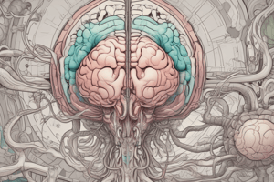
Basic Sectional and Radiographic Anatomy
- Skull comprises 8 cranial bones and 14 facial bones.
- Cranial bones are divided into calvaria (frontal, parietal, occipital) and base (temporal, sphenoid, ethmoid).
- Main skull sutures include coronal, sagittal, lambdoid, and squamosal; the metopic suture is sometimes present in adults.
- Cranial vault contains all intracranial structures; everything outside is classified as extracranial.
- Cranial fossae: anterior (frontal lobes), middle (temporal lobes), posterior (cerebellum and brain stem), and pituitary fossa (pituitary gland).
Sinuses
- Sinuses vary greatly among individuals, housing air and important for potential injuries.
- Maxillary sinuses are relevant for orbital injuries; sphenoid and ethmoid sinuses are involved in basal skull fractures.
- Mastoid air cells connect to the middle ear, while frontal sinuses can be absent in some individuals.
Meninges
- Meninges consist of three layers: dura mater, arachnoid membrane, and pia mater.
- Provide structural support for vasculature and protect the CNS with CSF from mechanical damage.
- Meningitis and intracranial bleeds often affect the meninges.
Dura Mater
- Thick and tough outermost layer of meninges, located beneath skull and vertebral bones.
- Contains dural venous sinuses responsible for venous drainage into internal jugular veins.
- Dural reflections include:
- Falx cerebri: separates the cerebral hemispheres.
- Tentorium cerebelli: separates occipital lobes from cerebellum with a midbrain passage.
- Falx cerebelli: separates cerebellar hemispheres.
- Diaphragma sellae: covers sphenoid's hypophysial fossa, allowing passage for the pituitary stalk.
Traumatic Brain Injury
- Intracranial hemorrhages include:
- Extra-axial: extradural, subdural, subarachnoid.
- Intra-axial hemorrhage results directly within brain tissues.
- Complications: mass effect, cerebral edema, herniation, and potential skull fractures.
Non-Traumatic Emergency Conditions
- Hydrocephalus: condition of excess cerebrospinal fluid.
- Infections such as meningitis, encephalitis, abscesses, and empyema pose serious risks.
- Acute ischemic stroke and hypoxic ischemic encephalopathy are critical conditions requiring immediate intervention.
Studying That Suits You
Use AI to generate personalized quizzes and flashcards to suit your learning preferences.