Podcast
Questions and Answers
Which of the following accurately describes the path of blood flow in the systemic circulation?
Which of the following accurately describes the path of blood flow in the systemic circulation?
- Blood flows from the right ventricle to the lungs and then back to the left atrium.
- Blood flows from the right atrium to the right ventricle, then to the body tissues, and finally back to the left atrium.
- Blood flows from the left ventricle to the aorta, then to the body tissues, and finally back to the right atrium. (correct)
- Blood flows from the left ventricle to the pulmonary artery, then to the lungs, and finally back to the right atrium.
Where is the apex of the heart located?
Where is the apex of the heart located?
- Anterior, inferior, and left. (correct)
- Posterior, superior, and right.
- Posterior, inferior, and left.
- Anterior, superior, and right.
Which layer of the pericardium is attached to the heart muscle?
Which layer of the pericardium is attached to the heart muscle?
- Pericardial cavity
- Visceral layer of the serous pericardium (epicardium) (correct)
- Fibrous pericardium
- Parietal layer of the serous pericardium
What is the function of the pericardial fluid?
What is the function of the pericardial fluid?
Which of the following describes the flow of blood through the pulmonary circulation?
Which of the following describes the flow of blood through the pulmonary circulation?
The ligamentum arteriosum is a remnant of what fetal structure?
The ligamentum arteriosum is a remnant of what fetal structure?
What is the main function of the fibrous pericardium?
What is the main function of the fibrous pericardium?
Which of the following is a potential consequence of inflammation of the pericardium (pericarditis)?
Which of the following is a potential consequence of inflammation of the pericardium (pericarditis)?
During isovolumetric relaxation, which valves are closed?
During isovolumetric relaxation, which valves are closed?
What causes the second heart sound (S2)?
What causes the second heart sound (S2)?
During diastole, what happens to the pressure in the ventricles?
During diastole, what happens to the pressure in the ventricles?
What is the main event that occurs during the rapid filling period of diastole?
What is the main event that occurs during the rapid filling period of diastole?
What is the significance of the incisura or notch in the aortic pressure tracing?
What is the significance of the incisura or notch in the aortic pressure tracing?
Which of these statements about atrial pressure waves is true?
Which of these statements about atrial pressure waves is true?
What is the approximate duration of the isovolumetric relaxation period?
What is the approximate duration of the isovolumetric relaxation period?
What percentage of ventricular filling is attributed to atrial contraction during diastole?
What percentage of ventricular filling is attributed to atrial contraction during diastole?
What represents the volume work of the heart?
What represents the volume work of the heart?
What is the relationship between the amount of blood filling the ventricles during diastole and the force of contraction?
What is the relationship between the amount of blood filling the ventricles during diastole and the force of contraction?
What factors contribute to preload?
What factors contribute to preload?
How does the Frank-Starling mechanism contribute to the heart's pumping ability?
How does the Frank-Starling mechanism contribute to the heart's pumping ability?
What is the optimal stretch of cardiac muscle fibers for maximum force of contraction?
What is the optimal stretch of cardiac muscle fibers for maximum force of contraction?
How does the Frank-Starling law equalize the output of both sides of the heart?
How does the Frank-Starling law equalize the output of both sides of the heart?
What is the relationship between cardiac reserve and heart disease?
What is the relationship between cardiac reserve and heart disease?
Besides preload, which other factors influence cardiac output?
Besides preload, which other factors influence cardiac output?
What physiological process is responsible for the transient redness observed on an arm after leaning on a hard surface?
What physiological process is responsible for the transient redness observed on an arm after leaning on a hard surface?
What is the primary function of nitric oxide (NO) in the vascular system?
What is the primary function of nitric oxide (NO) in the vascular system?
Which of the following is NOT an agonist of NO release?
Which of the following is NOT an agonist of NO release?
What is the primary role of bradykinin in the vascular system?
What is the primary role of bradykinin in the vascular system?
Which of the following substances is released from mast cells in injured tissue?
Which of the following substances is released from mast cells in injured tissue?
What is the main function of serotonin in the vascular system?
What is the main function of serotonin in the vascular system?
Which of the following is a long-term mechanism of blood flow regulation?
Which of the following is a long-term mechanism of blood flow regulation?
Nitroglycerin, a treatment for angina, achieves its therapeutic effect by:
Nitroglycerin, a treatment for angina, achieves its therapeutic effect by:
What is the primary function of capillaries?
What is the primary function of capillaries?
Which of the following is NOT a characteristic of arterioles?
Which of the following is NOT a characteristic of arterioles?
What is the term for the union of branches from two or more arteries, providing an alternative route for blood flow to a tissue?
What is the term for the union of branches from two or more arteries, providing an alternative route for blood flow to a tissue?
What is the name of the vessel that emerges from an arteriole and supplies a group of capillaries?
What is the name of the vessel that emerges from an arteriole and supplies a group of capillaries?
Which of the following tissues has a less extensive capillary network?
Which of the following tissues has a less extensive capillary network?
What is the primary mechanism by which arterioles regulate blood flow into capillaries?
What is the primary mechanism by which arterioles regulate blood flow into capillaries?
Which of the following is TRUE regarding anastomoses?
Which of the following is TRUE regarding anastomoses?
Which of the following factors contributes to resistance in blood vessels?
Which of the following factors contributes to resistance in blood vessels?
What is the effect of fluid compressing the heart?
What is the effect of fluid compressing the heart?
Which layer of the heart is responsible for its pumping action?
Which layer of the heart is responsible for its pumping action?
What is the function of the intercalated discs found in cardiac muscle fibers?
What is the function of the intercalated discs found in cardiac muscle fibers?
Which of the following is NOT a direct result of CPR?
Which of the following is NOT a direct result of CPR?
What anatomical feature allows for the effectiveness of CPR?
What anatomical feature allows for the effectiveness of CPR?
Flashcards
Systemic Circulation
Systemic Circulation
The left side of the heart pumps oxygenated blood to the body.
Pulmonary Circulation
Pulmonary Circulation
The right side of the heart pumps deoxygenated blood to the lungs.
Apex of the Heart
Apex of the Heart
The pointed end of the heart directed anteriorly and to the left.
Base of the Heart
Base of the Heart
Signup and view all the flashcards
Pericardium
Pericardium
Signup and view all the flashcards
Pericardial Cavity
Pericardial Cavity
Signup and view all the flashcards
Ligamentum Arteriosum
Ligamentum Arteriosum
Signup and view all the flashcards
Pericarditis
Pericarditis
Signup and view all the flashcards
Diastole
Diastole
Signup and view all the flashcards
Isovolumetric Relaxation
Isovolumetric Relaxation
Signup and view all the flashcards
Second Heart Sound (S2)
Second Heart Sound (S2)
Signup and view all the flashcards
Atrial Filling
Atrial Filling
Signup and view all the flashcards
Rapid Filling Phase
Rapid Filling Phase
Signup and view all the flashcards
Atrial Contraction
Atrial Contraction
Signup and view all the flashcards
Aortic Pressure Changes
Aortic Pressure Changes
Signup and view all the flashcards
Atrial Pressure Waves
Atrial Pressure Waves
Signup and view all the flashcards
Cardiac Tamponade
Cardiac Tamponade
Signup and view all the flashcards
Cardiopulmonary Resuscitation (CPR)
Cardiopulmonary Resuscitation (CPR)
Signup and view all the flashcards
Myocardium
Myocardium
Signup and view all the flashcards
Heart Chambers
Heart Chambers
Signup and view all the flashcards
Cardiac Reserve
Cardiac Reserve
Signup and view all the flashcards
Preload
Preload
Signup and view all the flashcards
Frank-Starling Law
Frank-Starling Law
Signup and view all the flashcards
End Diastolic Volume (EDV)
End Diastolic Volume (EDV)
Signup and view all the flashcards
Venous Return
Venous Return
Signup and view all the flashcards
Equal Output
Equal Output
Signup and view all the flashcards
Cardiac Contractility
Cardiac Contractility
Signup and view all the flashcards
Afterload
Afterload
Signup and view all the flashcards
Reactive Hyperemia
Reactive Hyperemia
Signup and view all the flashcards
Endothelium-derived Relaxing Factor
Endothelium-derived Relaxing Factor
Signup and view all the flashcards
Nitric Oxide (NO)
Nitric Oxide (NO)
Signup and view all the flashcards
Bradykinin
Bradykinin
Signup and view all the flashcards
Histamine
Histamine
Signup and view all the flashcards
Serotonin
Serotonin
Signup and view all the flashcards
Angiogenesis
Angiogenesis
Signup and view all the flashcards
Vasodilation
Vasodilation
Signup and view all the flashcards
Anastomosis
Anastomosis
Signup and view all the flashcards
Collateral circulation
Collateral circulation
Signup and view all the flashcards
Arterioles
Arterioles
Signup and view all the flashcards
Resistance vessels
Resistance vessels
Signup and view all the flashcards
Capillaries
Capillaries
Signup and view all the flashcards
Microcirculation
Microcirculation
Signup and view all the flashcards
Metarteriole
Metarteriole
Signup and view all the flashcards
Study Notes
Review of the Heart
- Blood circulates through two circuits: systemic and pulmonary.
- Systemic circulation pumps oxygenated blood from the left heart to the body.
- Pulmonary circulation pumps deoxygenated blood from the right heart to the lungs.
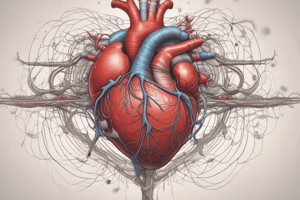
Anatomy of the Heart
- Located in the mediastinum, the central region of the thorax.
- Apex (pointed end) is directed anteriorly, inferiorly, and to the left.
- Base (opposite end) is directed posteriorly, superiorly, and to the right.
- Pericardium: A three-layered sac surrounding the heart composed of superficial fibrous and deeper serous pericardia (parietal and visceral layers); the space between the visceral and parietal layers is filled with fluid (pericardial fluid), to reduce friction.
Layers of the Heart Wall
- Epicardium: The outer layer of the heart wall, also the visceral layer of the serous pericardium.
- Myocardium: The thick, muscular middle layer, composed of cardiac muscle fibers forming atrial and ventricular networks. Intercalated discs (gap junctions and desmosomes) connect these fibers.
- Endocardium: The inner lining of the heart chambers, consisting of endothelium.
Heart Chambers
- Four chambers; two atria (receiving chambers) and two ventricles (pumping chambers).
- Right atrium receives deoxygenated blood from the body.
- Right ventricle pumps deoxygenated blood to the lungs.
- Left atrium receives oxygenated blood from the lungs.
- Left ventricle pumps oxygenated blood to the body.
Heart Valves
- Atrioventricular (AV) valves (tricuspid and mitral/bicuspid) prevent backflow from the ventricles to the atria during ventricular contraction.
- Semilunar valves (pulmonary and aortic) prevent backflow from the arteries into the ventricles during ventricular relaxation.
Cardiac Cycle
- One complete heartbeat, involving contraction (systole) and relaxation (diastole) of the atria and ventricles.
- Isovolumetric contraction: All valves closed, ventricular pressure increases.
- Ejection: Ventricular pressure exceeds arterial pressure, semilunar valves open.
- Isovolumetric relaxation: Ventricular pressure decreases, semilunar valves close.
- Ventricular filling: Atrial pressure exceeds ventricular pressure, AV valves open.
Cardiac Output
- Amount of blood pumped by the ventricles per minute.
- Determined by stroke volume (SV) and heart rate (HR).
- CO = SV × HR
Regulation of Cardiac Performance
- Cardiac output adjusts to meet the body's metabolic demands.
- Preload: Volume of blood in ventricles at the end of diastole.
- Afterload: Pressure ventricles must overcome to eject blood.
- Contractility: Strength of ventricular contraction.
- Heart rate: Influences cardiac output.
Blood Flow Through the Vessels
- Arteries: carry blood away from the heart.
- Arterioles: regulate blood flow into capillaries.
- Capillaries: sites of exchange between blood and tissues.
- Venules: collect blood from capillaries.
- Veins: carry blood toward the heart.
Venous Return
- Mechanisms for returning blood to the heart: skeletal muscle pump and respiratory pump.
- Pressure difference between the veins and atria drives venous return.
- An increase in right atrial or ventricular pressure can decrease venous return.
Studying That Suits You
Use AI to generate personalized quizzes and flashcards to suit your learning preferences.