Podcast
Questions and Answers
Wat is de functie van de discus articularis in het gewricht tussen het sternum en de clavicula?
Wat is de functie van de discus articularis in het gewricht tussen het sternum en de clavicula?
- Het maakt het gewricht gevoeliger voor blessures
- Het vergroot de mobiliteit van het gewricht
- Het zorgt voor aanpassingen omdat de gewrichtsvlakken zonder discus niet mooi in elkaar passen (correct)
- Het vermindert de stabiliteit van het gewricht
Welk type kraakbeen bedekt de gewrichtsvlakken van de articulatio sternoclavicularis?
Welk type kraakbeen bedekt de gewrichtsvlakken van de articulatio sternoclavicularis?
- Fibrocartilage
- Hyalien kraakbeen
- Elastisch kraakbeen
- Vezelkraakbeen (correct)
Welk ligament is verantwoordelijk voor het voorkomen van een daling van de laterale clavicula met meer dan 1 cm?
Welk ligament is verantwoordelijk voor het voorkomen van een daling van de laterale clavicula met meer dan 1 cm?
- Lig. Coracoclaviculare
- Lig. Sternoclaviculare posterius
- Lig. Interclaviculare
- Lig. Sternoclaviculare anterius (correct)
Wat is de functie van de incisura clavicularis van het sternum?
Wat is de functie van de incisura clavicularis van het sternum?
Wat is de functie van het radiale collaterale ligament aan de elleboog?
Wat is de functie van het radiale collaterale ligament aan de elleboog?
Wat wordt geremd door de articulatio humero-ulnaris?
Wat wordt geremd door de articulatio humero-ulnaris?
Wat is de functie van membrana interossea?
Wat is de functie van membrana interossea?
Wat kenmerkt de articulatio radio-ulnaris proximalis?
Wat kenmerkt de articulatio radio-ulnaris proximalis?
Wat is het doel van discus articularis in de articulatio radio-ulnaris distalis?
Wat is het doel van discus articularis in de articulatio radio-ulnaris distalis?
Wat veroorzaakt het remmen van adductie door lig. Anulare radii?
Wat veroorzaakt het remmen van adductie door lig. Anulare radii?
Welke beweging wordt geremd door de articulatio radio-ulnaris proximalis?
Welke beweging wordt geremd door de articulatio radio-ulnaris proximalis?
Wat is de maximale rotatie rond de as van de clavicula?
Wat is de maximale rotatie rond de as van de clavicula?
Hoeveel centimeter beweegt de clavicula naar boven bij cranio-caudale verplaatsing?
Hoeveel centimeter beweegt de clavicula naar boven bij cranio-caudale verplaatsing?
Wat is de maximale dorsoventrale verplaatsing van de clavicula?
Wat is de maximale dorsoventrale verplaatsing van de clavicula?
Wat voor type gewricht is het articulatio acromio-clavicularis?
Wat voor type gewricht is het articulatio acromio-clavicularis?
Hoe wordt de ligament Coracoclaviculair in de tekst beschreven?
Hoe wordt de ligament Coracoclaviculair in de tekst beschreven?
Wat is het effect van de verhoogde mobiliteit van de schoudergordel?
Wat is het effect van de verhoogde mobiliteit van de schoudergordel?
Wat kenmerkt de cavitas glenoidalis van de scapula?
Wat kenmerkt de cavitas glenoidalis van de scapula?
Hoe wordt de membrana fibrosa van capsula articularis in de tekst beschreven?
Hoe wordt de membrana fibrosa van capsula articularis in de tekst beschreven?
Wat kenmerkt labrum glenoidale volgens de tekst?
Wat kenmerkt labrum glenoidale volgens de tekst?
Wat is de functie van de ligamenta collateralia carpi?
Wat is de functie van de ligamenta collateralia carpi?
Welk bot vormt een scharniergewricht met het os trapezium en maakt oppositie van de duim mogelijk?
Welk bot vormt een scharniergewricht met het os trapezium en maakt oppositie van de duim mogelijk?
Welk type gewricht is de articulatio radiocarpale?
Welk type gewricht is de articulatio radiocarpale?
Wat kan er gebeuren bij fracturen aan het os scaphoideum aan de dorsale en radiale zijde?
Wat kan er gebeuren bij fracturen aan het os scaphoideum aan de dorsale en radiale zijde?
Hoe worden de ligamenten genoemd die het os pisiforme versterken?
Hoe worden de ligamenten genoemd die het os pisiforme versterken?
Wat is de functie van de ligamentum intercarpae?
Wat is de functie van de ligamentum intercarpae?
Welk type gewricht is de articulatio ossis pisiformis?
Welk type gewricht is de articulatio ossis pisiformis?
Wat is het belangrijkste bot voor beweging richting de handpalm in het polsgewricht?
Wat is het belangrijkste bot voor beweging richting de handpalm in het polsgewricht?
Wat vormt de facetten voor het gewrichtskapsels op het basis van de metacarpale botten?
Wat vormt de facetten voor het gewrichtskapsels op het basis van de metacarpale botten?
Wat wordt bedoeld met 'oppositie' van de duim?
Wat wordt bedoeld met 'oppositie' van de duim?
Wat is de primaire stabilisator van het mediale ellebooggewricht?
Wat is de primaire stabilisator van het mediale ellebooggewricht?
Welke functie heeft de voorste band van het ligamentum teres voor de humerus?
Welke functie heeft de voorste band van het ligamentum teres voor de humerus?
Waar bevindt zich de olecranonbursa?
Waar bevindt zich de olecranonbursa?
Welk effect hebben degeneratieve aandoeningen zoals osteoartritis op het ellebooggewricht?
Welk effect hebben degeneratieve aandoeningen zoals osteoartritis op het ellebooggewricht?
Hoe wordt het ellebooggewricht meestal behandeld bij letsel?
Hoe wordt het ellebooggewricht meestal behandeld bij letsel?
Wat is de rol van de articular cartilage in het ellebooggewricht?
Wat is de rol van de articular cartilage in het ellebooggewricht?
Welke structuur bevindt zich tussen het synoviale membraan en het fibreuze membraan in het ellebooggewricht?
Welke structuur bevindt zich tussen het synoviale membraan en het fibreuze membraan in het ellebooggewricht?
Waar bevindt zich de ulnar nerve in relatie tot het ellebooggewricht?
Waar bevindt zich de ulnar nerve in relatie tot het ellebooggewricht?
Wat is de functie van ligamentum anulare in het ellebooggewricht?
Wat is de functie van ligamentum anulare in het ellebooggewricht?
Hoeveel functionele gewrichten bevat het ellebooggewricht binnen één gewrichtscapsule?
Hoeveel functionele gewrichten bevat het ellebooggewricht binnen één gewrichtscapsule?
Flashcards are hidden until you start studying
Study Notes
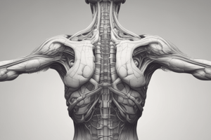
- Articulatio humeri (shoulder joint): ball-and-socket joint with center in head, limited abduction to 90° due to tuberculum majus and coraco-acromial ligament. Elevation requires involvement of other shoulder girdle joints. Adduction is restricted by body. Light antiflexion allows for only 30 degrees of adduction. Anteflexion is not limited by ligaments and can exceed 90 degrees of elevation. Retroflexion is strongly limited by glenohumeral ligaments in the front of the joint capsule. Exorotation (80°) and endorotation (100°) are limited by the capsule. Protractio and retractie can only occur when both other joints are involved. Glenohumeral joint is less mobile than hip joint. Shoulder girdle is more mobile due to two additional joints (sternoclavicularis and acromioclavicularis) that pull the shoulder blade forward and back. Increased mobility comes at the cost of stability.
- The center of pure abduction in the glenohumeral joint is 90° to 120°, but a limited lowering of the humerus head during this movement must be considered.
- The combination of ab- and adductiebewegingen (all ab- and adductive movements), ante- and retroflexies with an extended arm describes a line within the realm of the spherical surface. The center of this surface is the head of the humerus. At each point in the excursion range, the limb can describe rotation. When the joint becomes stiff, remaining mobility of the shoulder girdle (sternoclavicularis and acromioclavicularis joints) is preserved. This kind of stiffness can occur after fractures, luxations, or chronic shoulder pain and is a result of capsule retractive (scarring).
- The global shoulder girdle functions passively with the skeleton and its ligamentous bands carrying the arm without the intervention of muscles. The clavicle is horizontally fixed by the sternoclavicular joint, and the scapula hangs from the clavicle by the conoidal and trapezoid ligaments. The scapula rests against the 7th rib and has a 7-degree space underneath.
- The humerus is fixed posteriorly by muscles and anteriorly by ligaments. Actively, in each standing position of the glenoid cavity that is allowed by the clavicular joints, the articulatio humeri (shoulder joint) can describe its excursion range. Through changes in the position of the glenoid cavity, an excursion space is created. At each point in this space, rotation of the arm is possible. With each shoulder movement, all the joints in the system come into play, including those that could have been involved in a single joint movement alone.
- The elbow joint: three functional joints within one joint capsule. The articular surfaces are all covered with hyaline cartilage. 1. Articulatio humero-ulnaris (ulno-humeral joint): hinge joint. On the ulna: trochlea. On the humerus: trochlea. 2. Articulatio humero-radialis (radio-humeral joint): spherical joint. Humerus: round head. Radius: articulating surface: fovea on the head of the radius. 3. Articulatio radio-ulnaris proximalis (proximal radioulnar joint): pivot joint. Radius: circumferential articular surface. Ulna: radial notch. The number of joint axes equals 5. The joint capsule is common for the three joints. The fibrous membrane is abnormally attached to the humerus. The fossa olecrani, radialis, and coronoidea ligaments are found infra-articularly, as are the condyles of the humerus consisting of the trochlea, capitulum, and three fossae.
- The epicondyles remain free for muscle insertions. On the ulna, the attachment is almost normal at the bone edge. The olecranon and processus conoideus are still in the joint capsule. On the radius, the attachment is normal, but just distal from the border of the circumferential articular surface. This forms a sac-like pouch around the radius. The elbow joint is a hinge joint (with three parts). Between the synovial membrane and the fibrous membrane lies fatty tissue that has a dampening effect.
- The ligaments play a crucial role in the elbow joint: 1. Ligamentum anulare: this ligament holds the radius to the ulna in every position and only allows rotation along the long axis; it is centered in the circumferential ligament. 2. Ligamentum quadraturum: this ligament forms reinforcement of the sac-like pouch in the space between the radius and ulna. 3. Transverse bands run from the distal edge of the radial notch to the ulna-side of the collarbone. Close to the attachment on the ulna, circular fibers pass through the transverse bands. These fibers can be considered as the distal part of the ligamentum anulare. 4. Ligamentum collaterale ulnare: this broad triangular band is firmly fixed to the epicondyle medialis, with the front part consisting of strong parallel fibers that run to the ulna's radial side of the processus coronoideus, and the rear part runs to the ulna's side of the olecranon. Pars transversa connects both parts distally with transverse bundles. Function: it limits abduction; the front part checks extension, and the posterior part checks flexion.
- The ulnar nerve runs posterior to the elbow joint, and the radial nerve runs anterior to it. The brachial artery runs anterior to the elbow joint, and the posterior interosseous artery runs posterior to it. The ulnar artery and the radial artery form the anastomosis between the elbow and the distal forearm.
- In the elbow joint, the articular cartilage is thicker in the medial compartment than in the lateral compartment due to the greater load experienced by the medial compartment. The ligaments are less elastic on the medial side than on the lateral side.
- The ulnar collateral ligament is the primary stabilizer of the medial elbow joint, while the lateral collateral ligament provides stability in flexion and extension.
- The anterior band of the ligamentum teres functions as a rotator cuff for the humerus.
- The olecranon bursa is a synovial-lined sac that lies between the olecranon and the triceps muscle. It is a common site for bursitis.
- The elbow is a complex joint with a large number of muscles, ligaments, and nerves involved in its function. It is essential for movements such as flexion, extension, supination, and pronation of the forearm.
- The elbow joint is also involved in the transmission of forces during activities such as lifting, pushing, and pulling.
- The elbow joint is prone to injuries, including fractures, ligament ruptures, and dislocations. These injuries can result in chronic pain and loss of function if not treated promptly and properly.
- The elbow joint is susceptible to degenerative conditions such as osteoarthritis, which can cause pain and stiffness.
- The elbow joint is commonly used in daily life, and its health and functionality are crucial for maintaining a good quality of life.
- The elbow joint is the second most commonly used joint in the body, after the shoulder joint.
- The elbow joint is a synovial joint with a fibrocartilaginous meniscus in the trochlea, which provides stability and absorbs shock.
- The elbow joint is the most mobile joint in the upper limb, allowing for a wide range of motion.
- The elbow joint is often treated with conservative measures such as rest, ice, compression, and elevation, as well as physical therapy and medication.
- The elbow joint can also be treated with surgical intervention, such as arthroscopy or joint replacement, in cases of severe injury or degenerative conditions.
- The elbow joint is important for activities such as writing, playing musical instruments, and using tools, making its health and functionality crucial for various aspects of daily life.
- The elbow joint is a complex joint with a large number of muscles, ligaments, and nerves involved in its function. It is essential for movements such as flexion, extension, supination, and pronation of the forearm. It is also involved in the transmission of forces during activities such as lifting, pushing, and pulling.
- The elbow joint is prone to injuries, including fractures, ligament ruptures
Studying That Suits You
Use AI to generate personalized quizzes and flashcards to suit your learning preferences.


