Podcast
Questions and Answers
Adapta le sequente musculos con su location:
Adapta le sequente musculos con su location:
Tibialis Anterior = Gambe Gastrocnemius = Gambe Peroneus Longus = Gambe Peroneus Brevis = Gambe Soleus = Gambe Extensor Digitorium Longus = Gambe Extensor Hallucis Longus = Gambe
Quale de iste optiones es le partes del corde?
Quale de iste optiones es le partes del corde?
- Dextre Ventriculo (correct)
- Sinistre Atrium (correct)
- Dextre Atrium (correct)
- Sinistre Ventriculo (correct)
- Pericardium (correct)
Le ______ es le parte del musculo que contrahe.
Le ______ es le parte del musculo que contrahe.
ventre
Adapta le sequente oses con su location:
Adapta le sequente oses con su location:
Adapta le sequente arterias con su location:
Adapta le sequente arterias con su location:
Adapta le sequente partes del cella con su description:
Adapta le sequente partes del cella con su description:
Flashcards
Tibia
Tibia
Le tibia es un oss del gamba que es situate in le parte inferior del gamba.
Fibula
Fibula
Le fibula es un oss del gamba que es situate in le parte inferior del gamba.
Patella
Patella
Le patella es un oss que es situate in le genuo.
Femure
Femure
Signup and view all the flashcards
Talus
Talus
Signup and view all the flashcards
Tarsos
Tarsos
Signup and view all the flashcards
Metatarsos
Metatarsos
Signup and view all the flashcards
Phalanges
Phalanges
Signup and view all the flashcards
Humerus
Humerus
Signup and view all the flashcards
Radius
Radius
Signup and view all the flashcards
Ulna
Ulna
Signup and view all the flashcards
Lunate
Lunate
Signup and view all the flashcards
Metacarpos
Metacarpos
Signup and view all the flashcards
Phalanges
Phalanges
Signup and view all the flashcards
Pericardium
Pericardium
Signup and view all the flashcards
Atrio Dextre
Atrio Dextre
Signup and view all the flashcards
Atrio Sinistre
Atrio Sinistre
Signup and view all the flashcards
Ventriculo Dextre
Ventriculo Dextre
Signup and view all the flashcards
Ventriculo Sinistre
Ventriculo Sinistre
Signup and view all the flashcards
Study Notes
Anatomical Structures: Lower Limb
- Tibialis Anterior: Muscle in the lower leg, involved in foot movement.
- Gastrocnemius: Muscle in the calf, crucial for plantar flexion.
- Peroneus Longus: Muscle contributing to foot eversion and plantarflexion.
- Peroneus Brevis: Muscle involved in foot eversion and plantarflexion.
- Soleus: Muscle of the lower leg, responsible for plantar flexion.
- Extensor Digitorum Longus: Muscle that extends toes and dorsiflexes the foot.
- Extensor Hallucis Longus: Extends the big toe and dorsiflexes the foot.
- Flexor Digitorum Brevis: Flexes the smaller toes.
- Abductor Hallucis: Abducts the big toe.
- Abductor Digiti Minimi: Abducts the little toe.
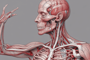
Anatomical Structures: Circulatory System
- Pericardium: The sac surrounding the heart.
- Right Atrium: Receives deoxygenated blood from the body.
- Left Atrium: Receives oxygenated blood from the lungs.
- Right Ventricle: Pumps deoxygenated blood to the lungs.
- Left Ventricle: Pumps oxygenated blood to the body.
Anatomical Structures: Upper Limb
- Origin (muscle): The part of a muscle where it attaches to bone.
- Belly (muscle): The fleshy part between origin and insertion.
- Insertion (muscle): Where the muscle attaches to another stable bone.
- Humerus: Bone of the upper arm.
- Radius: A bone of the forearm.
- Ulna: A bone of the forearm.
- Carpals: Small bones forming the wrist.
- Metacarpals: Bones in the hand, located between wrist and fingers
- Phalanges: Bones forming the fingers.
- Patella: Knee cap.
- Tibia: Shin bone.
- Fibula: Smaller bone in the lower leg.
- Talus: Ankle bone
Anatomical Structures: Head and Neck
- Frontalis: Raising eyebrows.
- Orbicularis Oculi: Muscle around the eye for closing.
- Orbicularis Oris: Muscle around the mouth.
- Mentalis: Raises the lower lip
- **Zygomaticus:**Raises the corners of the mouth
- Caninus: Draws corners of mouth upward and backward.
- Buccinator: Compresses the cheek
- Corrugator Supercilii: Draws eyebrows together
- Levator Palpebrae Superioris: Raises the upper eyelid
Anatomical Structures: Ear
- Auricularis Anterior (muscle): Draws the ear forward
- Auricularis Superior(muscle): Draws the ear upward
- Auricularis Posterior (muscle): Draws the ear back
Anatomical Structures: Nervous System
- Nerve Cell (Neuron): The basic unit of the nervous system.
- Axon: Transmits nerve impulses away from the cell body.
- Dendrites: Receive signals from other neurons.
- Ulnar: Nerve that supplies part of the forearm and hand.
- Radial: Nerve that supplies part of the arm and forearm.
- Median: Nerve supplying the palm, and some fingers.
- Digital: Nerves of the fingers
- Sciatic: Main nerve of the buttock and leg
- Anterior tibial: Nerve that supplies front of the lower leg and foot
- Posterior tibial: Nerve that supplies posterior part of the foot
- Common peroneal: Nerve that supplies lateral side of the leg and foot
- Deep peroneal: Nerve that supplies muscles and skin on anterior of foot
- Superficial peroneal: Nerve that supplies muscles and skin on lateral side of foot
- Saphenous: nerve that supplies skin on medial side of leg
- Sural: nerve that supplies posterior and lateral side of foot and leg
- Popliteal: Main artery in the back of the knee
- Dorsalis pedis: Artery in the foot
- Femoral: Artery that branches from the aorta and supplies the thigh region.
Anatomical Structures: Skull
- Mandible: Jaw bone.
- Maxilla: Upper jaw bone.
- Nasal: Bones forming the bridge of the nose.
- Zygomatic: Cheek bone.
- Lacrimal: Small bone next to the eye socket.
- Frontal: Forehead bone.
- Parietal: Bones forming the sides and top of the skull.
- Occipital: Back of the skull.
- Temporal: Bones on the sides of the skull.
Anatomical Structures: Spinal Column
- Vertebrae: Bones of the spinal column.
Anatomical Structures: Thorax
- Ribs: Bones that form the rib cage protecting the thoracic cavity and lungs.
- Sternum: Breastbone, also part of thorax structure.
Anatomical Structures: Musculature
- Pectoralis Major: Large chest muscle.
- Pectoralis Minor: Smaller, underlying chest muscle.
- Serratus Anterior: Muscle in the side of the chest, responsible for winging the scapula.
- Deltoid: Shoulder muscle
- Biceps: Muscle in the front of the upper arm.
- Triceps: Muscle in the back of the upper arm.
- Supinator: Muscle that supports the forearm.
- Pronator: Muscle that pronates the forearm.
- Flexor: Muscle involved in bending
- Extensor: Muscle involved in straightening the body part.
- Abductor: Muscle responsible for moving a part away from the body.
- Adductor: Muscle responsible for moving a part towards the body.
- Opponens: Muscle responsible for flexing
Anatomical Structures: Cell Biology
- Nucleus: Control center of the cell.
- Cytoplasm: Substance filling the cell.
- Cell Membrane: Outer boundary of the cell.
Other Anatomical Structures
- Femur (bone): Thigh bone.
- Tarsals (bones): Forming the ankle structure.
- Metatarsals (bones): Between the tarsus and phalanges
- Phalanges (bones): Part of the digits of the hands and feet
Studying That Suits You
Use AI to generate personalized quizzes and flashcards to suit your learning preferences.