Podcast
Questions and Answers
¿Qué función relacionada con el tacto está asociada a las fibras simpáticas?
¿Qué función relacionada con el tacto está asociada a las fibras simpáticas?
- Movimientos reflejos
- Vibración (correct)
- Percepción del frío
- Sensación de presión
¿Cuál de las siguientes estructuras está relacionada con el nervio accesorio?
¿Cuál de las siguientes estructuras está relacionada con el nervio accesorio?
- Trapecio (correct)
- Esternocleidomastoideo (correct)
- Pectoral mayor
- Deltoides
¿Qué tipo de información proporcionan las aferencias viscerales?
¿Qué tipo de información proporcionan las aferencias viscerales?
- Información sobre cambios químicos (correct)
- Sensaciones dolorosas
- Sensaciones táctiles
- Información sobre equilibrio
¿Cuál de los siguientes tipos de fibras NO está asociado al epicrítico?
¿Cuál de los siguientes tipos de fibras NO está asociado al epicrítico?
¿Qué nervios son afectados por neuronas preganglionares en el sistema simpático?
¿Qué nervios son afectados por neuronas preganglionares en el sistema simpático?
¿Qué tipo de sensación está asociada al movimiento en el contexto de las fibras simpáticas?
¿Qué tipo de sensación está asociada al movimiento en el contexto de las fibras simpáticas?
¿Qué nervio se asocia principalmente con información aferente visceral?
¿Qué nervio se asocia principalmente con información aferente visceral?
¿Cuál es la principal característica de las fibras preganglionares en las vías simpáticas?
¿Cuál es la principal característica de las fibras preganglionares en las vías simpáticas?
¿Cuál de los nervios craneales está relacionado con la audición y el equilibrio?
¿Cuál de los nervios craneales está relacionado con la audición y el equilibrio?
¿Qué función tiene el nervio craneal NC III?
¿Qué función tiene el nervio craneal NC III?
¿Qué estructura se asocia con el nervio craneal NC IX?
¿Qué estructura se asocia con el nervio craneal NC IX?
¿Cuál de los siguientes nervios craneales tiene funciones parasimpáticas?
¿Cuál de los siguientes nervios craneales tiene funciones parasimpáticas?
¿Qué músculo es inervado por el nervio craneal NC XI?
¿Qué músculo es inervado por el nervio craneal NC XI?
¿Qué nervio está involucrado en la percepción del gusto?
¿Qué nervio está involucrado en la percepción del gusto?
¿Qué función realiza el nervio NC IV?
¿Qué función realiza el nervio NC IV?
¿Cuál de los siguientes nervios está relacionado con la laringe?
¿Cuál de los siguientes nervios está relacionado con la laringe?
¿Cuál es la parte superior del mesencéfalo?
¿Cuál es la parte superior del mesencéfalo?
¿Qué nervio se encuentra debajo del colículo inferior en el mesencéfalo?
¿Qué nervio se encuentra debajo del colículo inferior en el mesencéfalo?
¿Cuál es el límite inferior del puente?
¿Cuál es el límite inferior del puente?
¿Qué estructura marca el límite superior del puente?
¿Qué estructura marca el límite superior del puente?
¿Cuál es la función principal del cono medular?
¿Cuál es la función principal del cono medular?
¿Qué limite inferior define al bulbo raquídeo?
¿Qué limite inferior define al bulbo raquídeo?
¿Dónde se localiza el fillum terminal?
¿Dónde se localiza el fillum terminal?
¿Cuál es el límite superior del mesencéfalo?
¿Cuál es el límite superior del mesencéfalo?
¿Qué estructura está relacionada con la cauda equina?
¿Qué estructura está relacionada con la cauda equina?
Entre cuál y cuál estructuras se ubica el puente?
Entre cuál y cuál estructuras se ubica el puente?
¿Qué es el fillum dentado?
¿Qué es el fillum dentado?
¿Cuál es la ubicación del final de la duramadre?
¿Cuál es la ubicación del final de la duramadre?
¿Qué caracteriza al plexo coroideo en el contexto del LCR?
¿Qué caracteriza al plexo coroideo en el contexto del LCR?
¿Qué liga al fillum terminal con la médula espinal?
¿Qué liga al fillum terminal con la médula espinal?
¿Cuál es el principal componente del sistema nervioso central mencionado?
¿Cuál es el principal componente del sistema nervioso central mencionado?
¿Qué función tienen las raíces lumbosacras en la cauda equina?
¿Qué función tienen las raíces lumbosacras en la cauda equina?
¿Cuál es la consecuencia de una lesión en el nervio cráneo III (NC III)?
¿Cuál es la consecuencia de una lesión en el nervio cráneo III (NC III)?
La miosis se produce cuando ocurre la contracción de:
La miosis se produce cuando ocurre la contracción de:
Una parálisis del nervio cráneo IV (NC IV) puede resultar en:
Una parálisis del nervio cráneo IV (NC IV) puede resultar en:
¿Qué tipo de respuesta se espera de la pupila durante la midriasis?
¿Qué tipo de respuesta se espera de la pupila durante la midriasis?
La función del músculo elevador del párpado está relacionada principalmente con:
La función del músculo elevador del párpado está relacionada principalmente con:
¿Cuál de los siguientes músculos se contrae para producir miosis?
¿Cuál de los siguientes músculos se contrae para producir miosis?
Una lesión en el nervio facial (NC VII) puede causar:
Una lesión en el nervio facial (NC VII) puede causar:
El músculo que ayuda en la rotación del ojo hacia abajo y afuera es:
El músculo que ayuda en la rotación del ojo hacia abajo y afuera es:
¿Cuál de las siguientes estructuras está implicada en la sensibilidad facial?
¿Cuál de las siguientes estructuras está implicada en la sensibilidad facial?
¿Qué tipo de fibras conecta diferentes áreas dentro del mismo hemisferio cerebral?
¿Qué tipo de fibras conecta diferentes áreas dentro del mismo hemisferio cerebral?
¿Cuál es la función principal del músculo estilogloso?
¿Cuál es la función principal del músculo estilogloso?
¿Cuál de las siguientes opciones describe mejor las fibras comisurales?
¿Cuál de las siguientes opciones describe mejor las fibras comisurales?
¿Cuál nervio está encargado de la movilidad de la mandíbula?
¿Cuál nervio está encargado de la movilidad de la mandíbula?
¿Qué función cumplen las fibras de proyección?
¿Qué función cumplen las fibras de proyección?
¿Qué estructura se considera parte de las fibras en U?
¿Qué estructura se considera parte de las fibras en U?
¿Cuál de los siguientes nervios está involucrado en el movimiento del músculo palatogloso?
¿Cuál de los siguientes nervios está involucrado en el movimiento del músculo palatogloso?
Flashcards
¿Qué es la cola de caballo?
¿Qué es la cola de caballo?
Parte final de las raíces nerviosas lumbares y sacras que descienden hacia el canal vertebral.
¿Qué es el cono medular?
¿Qué es el cono medular?
La parte final de la médula espinal, que se estrecha.
¿Qué es el filum terminal?
¿Qué es el filum terminal?
Una prolongación de la piamadre que desciende por debajo del cono medular.
¿Dónde termina la médula espinal?
¿Dónde termina la médula espinal?
Signup and view all the flashcards
Función del cono medular
Función del cono medular
Signup and view all the flashcards
Localización de la cola de caballo
Localización de la cola de caballo
Signup and view all the flashcards
Estructura del filum terminal
Estructura del filum terminal
Signup and view all the flashcards
Función del filum terminal
Función del filum terminal
Signup and view all the flashcards
Fibras simpáticas
Fibras simpáticas
Signup and view all the flashcards
Tacto epicrítico
Tacto epicrítico
Signup and view all the flashcards
Movimiento
Movimiento
Signup and view all the flashcards
Vibración
Vibración
Signup and view all the flashcards
Nervio accesorio
Nervio accesorio
Signup and view all the flashcards
Pre-ganglionares
Pre-ganglionares
Signup and view all the flashcards
Núcleo aferente visceral
Núcleo aferente visceral
Signup and view all the flashcards
Información aferente visceral
Información aferente visceral
Signup and view all the flashcards
Miosis
Miosis
Signup and view all the flashcards
Midriasis
Midriasis
Signup and view all the flashcards
¿Qué músculos controlan el tamaño de la pupila?
¿Qué músculos controlan el tamaño de la pupila?
Signup and view all the flashcards
Parálisis del III par craneal (NC III)
Parálisis del III par craneal (NC III)
Signup and view all the flashcards
Parálisis del IV par craneal (NC IV)
Parálisis del IV par craneal (NC IV)
Signup and view all the flashcards
Parálisis del VI par craneal (NC VI)
Parálisis del VI par craneal (NC VI)
Signup and view all the flashcards
¿Cuáles son las consecuencias de una lesión al NC III?
¿Cuáles son las consecuencias de una lesión al NC III?
Signup and view all the flashcards
¿Cuáles son las consecuencias de una lesión al NC IV?
¿Cuáles son las consecuencias de una lesión al NC IV?
Signup and view all the flashcards
Tallo Cerebral
Tallo Cerebral
Signup and view all the flashcards
Mesencéfalo
Mesencéfalo
Signup and view all the flashcards
Límites del Mesencéfalo
Límites del Mesencéfalo
Signup and view all the flashcards
Nervios del Mesencéfalo
Nervios del Mesencéfalo
Signup and view all the flashcards
Puente
Puente
Signup and view all the flashcards
Límites del Puente
Límites del Puente
Signup and view all the flashcards
Bulbo Raquídeo
Bulbo Raquídeo
Signup and view all the flashcards
Límites del Bulbo Raquídeo
Límites del Bulbo Raquídeo
Signup and view all the flashcards
NC VIII
NC VIII
Signup and view all the flashcards
NC IX
NC IX
Signup and view all the flashcards
NC X
NC X
Signup and view all the flashcards
NC XI
NC XI
Signup and view all the flashcards
NC III
NC III
Signup and view all the flashcards
NC IV
NC IV
Signup and view all the flashcards
¿Qué función tiene la glándula parótida?
¿Qué función tiene la glándula parótida?
Signup and view all the flashcards
¿Qué funciones realiza el nervio accesorio (NC XI)?
¿Qué funciones realiza el nervio accesorio (NC XI)?
Signup and view all the flashcards
Fibras Comisurales
Fibras Comisurales
Signup and view all the flashcards
Fibras de Asociación
Fibras de Asociación
Signup and view all the flashcards
Fibras de Proyección
Fibras de Proyección
Signup and view all the flashcards
Cuerpo Calloso
Cuerpo Calloso
Signup and view all the flashcards
Paladar
Paladar
Signup and view all the flashcards
Faringe y Laringe
Faringe y Laringe
Signup and view all the flashcards
Músculo Palatogloso
Músculo Palatogloso
Signup and view all the flashcards
Nervios Craneales
Nervios Craneales
Signup and view all the flashcards
Study Notes
General Anatomy
- The body is composed of many parts working together, including the central nervous system (CNS) and the peripheral nervous system (PNS).
- The CNS includes the brain and spinal cord.
- The PNS extends outwards from the CNS and includes the cranial nerves and spinal nerves.
Spinal Cord
- The spinal cord's superior limit is the foramen magnum and the pyramidal decussation.
- Its inferior boundary is at L1-L2, or L3 in children.
- It runs through the vertebral foramen.
- Anteriorly, it is marked by the anterior median fissure.
- Posteriorly, it is marked by the posterior median sulcus.
- It is composed of 31 segments (including nerves C1-C3), with varying sizes.
- Its approximate length ranges from 43-45cm.
- The spinal cord is enclosed by meninges (membranes).
- Cerebrospinal fluid (CSF) circulates within the subarachnoid space acting as a cushion.
- Cervical and lumbar enlargements are present, yielding the brachial and lumbar plexuses respectively
- The cauda equina is the collection of nerve roots extending below the medullary cone
- The medullary cone is the tapered end of spinal cord
- The filum terminale is an extension of the pia mater that anchors the spinal cord inferiorly.
- The spinal cord receives blood supply from branches of the vertebral and subclavian arteries.
- Gray matter is centrally located, comprising neuron cell bodies and unmyelinated axons.
- White matter is externally situated, comprised myelinated axons.
- Afferent neurons are found in posterior horns, carrying sensory info.
- Efferent neurons are in anterior horns, conveying motor signals.
Brain
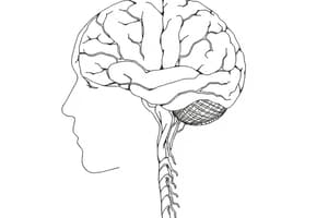
- The brain comprises the cerebrum (diencephalon + telencephalon), midbrain, cerebellum, and brainstem (pons and medulla).
- The cerebrum is divided into lobes (frontal, parietal, temporal, occipital).
- Several grooves and gyri are present on the surface
Ventricles of the Brain
- The cerebral ventricles are four fluid-filled spaces within the brain, filled with cerebrospinal fluid (CSF).
- The Lateral Ventricles (2) communicate with the third ventricle through the interventricular foramina.
- The third ventricle is found below the lateral ventricles and also below the cerebral aqueduct (mesencephalic).
- The fourth ventricle is continuous with the central canal of the spinal cord, and continues posteriorly through foramina (openings) to the subarachnoid space.
- The choroid plexuses in ventricles produce CSF at a rate of ~0.5 mL/ minute (~500 mL/24 hr) via the blood vessels.
Brainstem:
- The brainstem is comprised of the midbrain, pons, and medulla.
- Cranial nerves exit the brainstem, with various functional roles (motor, sensory, or both)
- It plays a critical role in regulating vital functions like breathing, heart rate, and consciousness.
Cerebellum
- The cerebellum is a structure located posteriorly to the brainstem.
- The cerebellum plays a significant role in coordinating movement and maintaining balance
- The cerebellum has distinct lobes (anterior, posterior, and flocculonodular).
- The key nuclei are the fastigial, dentate, globose, and emboliform nuclei.
Cranial Nerves
- The cranial nerves (12 pairs) have various sensory, motor, or mixed duties,
- Including vision, smell, taste, eye movement, facial expression, hearing, swallowing, and others.
- Their functions range for conveying sensory information from parts of the body.
Peripheral Nervous System
- The PNS includes the cranial and spinal nerves, extending from the CNS.
Pelvis and Gluteal Region
- The area is bordered by the iliac crest, greater trochanter, and sacrum.
- Bones (ilium, ischium, pubis) make up the pelvis.
- Muscles like the gluteus maximus, medius, and minimus are responsible for hip extension, abduction, and medial rotation.
- Other muscles (piriformis, obturator internus, and gemelli, quadratus femoris) are deep, primarily responsible for lateral rotation -Key ligaments (iliofemoral, pubofemoral, ischiofemoral, transverse acetabular and ligament of the head of the femur) help stabilize the hip
- The region contains important structures like the femoral artery and nerve.
Femoral Region
- The femoral region extends from the groin to the knee.
- The major anterior muscles are the quadriceps femoris (rectus femoris, vastus lateralis, vastus medialis, and vastus intermedius), hip flexors (iliopsoas, sartorius), and adductors (gracilis, adductor longus, adductor magnus).
- The femoral artery is a significant vessel in this region.
Knee Joint
- The knee is a complex hinge joint with ligaments (cruciate, collateral, menisci) playing roles in stability.
- The knee joint, and associated structures, can be subject damage by a variety of forces.
Leg and Ankle
- The tibia and fibula form the leg's structural framework .
- The major muscles (tibialis anterior, extensor digitorum longus, fibularis longus, gastrocnemius, soleus, tibialis posterior, flexor digitorum longus, flexor hallucis longus) affect movement.
- The ankle is a joint with ligaments stabilizing ankle mobility.
- Structures like tendons and arteries provide support & function in the lower leg and ankle
Foot
- The foot features tarsal and metatarsal bones.
- The arrangement of muscles in the foot's sole permits intricate actions.
Studying That Suits You
Use AI to generate personalized quizzes and flashcards to suit your learning preferences.


