Podcast
Questions and Answers
What do the Striae of Retzius represent in enamel structure?
What do the Striae of Retzius represent in enamel structure?
- The presence of enamel tufts
- Concentric rings of dentin
- The transition from prenatal to postnatal enamel
- Incremental lines of enamel formation (correct)
Where is the neonatal line primarily observed?
Where is the neonatal line primarily observed?
- In the cervical region of the tooth
- Only in permanent molars
- In all deciduous teeth and first permanent molars (correct)
- In the incisal edge of incisors only
Which type of enamel lamellae is characterized by poorly calcified enamel rods?
Which type of enamel lamellae is characterized by poorly calcified enamel rods?
- Type D
- Type C
- Type A (correct)
- Type B
What are enamel tufts primarily composed of?
What are enamel tufts primarily composed of?
How far do enamel spindles typically extend into the enamel?
How far do enamel spindles typically extend into the enamel?
What distinguishes the enamel lamellae from enamel tufts?
What distinguishes the enamel lamellae from enamel tufts?
What causes the neonatal line to become prominent?
What causes the neonatal line to become prominent?
What is a characteristic of Type C enamel lamellae?
What is a characteristic of Type C enamel lamellae?
What is the primary function of the reduced enamel epithelium?
What is the primary function of the reduced enamel epithelium?
Which type of ameloblast is more prevalent during the maturation proper phase?
Which type of ameloblast is more prevalent during the maturation proper phase?
How is enamel structurally characterized?
How is enamel structurally characterized?
What can result if connective tissue comes into contact with enamel?
What can result if connective tissue comes into contact with enamel?
What is the composition of enamel rods?
What is the composition of enamel rods?
What is the significance of the dark lines seen in longitudinal sections of enamel rods?
What is the significance of the dark lines seen in longitudinal sections of enamel rods?
What happens to the epithelial cells during the desmolytic stage?
What happens to the epithelial cells during the desmolytic stage?
What is a unique property of enamel compared to other calcified tissues in the body?
What is a unique property of enamel compared to other calcified tissues in the body?
What is one of the main components of dentin?
What is one of the main components of dentin?
Which statement accurately describes the dentinal tubules?
Which statement accurately describes the dentinal tubules?
What is the primary characteristic of Hunter-Schreger bands?
What is the primary characteristic of Hunter-Schreger bands?
What function do odontoblast processes serve?
What function do odontoblast processes serve?
What distinguishes gnarled enamel from regular enamel?
What distinguishes gnarled enamel from regular enamel?
What is the origin of odontoblasts?
What is the origin of odontoblasts?
What is the first phase in the process of dentinogenesis?
What is the first phase in the process of dentinogenesis?
How do the odontoblastic processes relate to the dentinal tubules?
How do the odontoblastic processes relate to the dentinal tubules?
Which type of dentin is formed after root completion?
Which type of dentin is formed after root completion?
What characterizes tertiary dentin?
What characterizes tertiary dentin?
Which of the following describes simple mantle dentin?
Which of the following describes simple mantle dentin?
What is NOT a characteristic of primary dentin?
What is NOT a characteristic of primary dentin?
What term is used for the unmineralized layer of dentin deposited first during formation?
What term is used for the unmineralized layer of dentin deposited first during formation?
What type of dentin acts as a barrier within dentinal tubules?
What type of dentin acts as a barrier within dentinal tubules?
Which characteristic is true regarding intertubular dentin?
Which characteristic is true regarding intertubular dentin?
What signifies the transition between prenatal and postnatal dentin?
What signifies the transition between prenatal and postnatal dentin?
Which type of dentin is characterized by calcification in the dentinal tubules?
Which type of dentin is characterized by calcification in the dentinal tubules?
Incremental lines of Von Ebner in dentin are primarily associated with what?
Incremental lines of Von Ebner in dentin are primarily associated with what?
What occurs in dentin as a result of aging or injury?
What occurs in dentin as a result of aging or injury?
The zone of hypo-mineralized dentin separating mantle dentin and circumpulpal dentin is known as?
The zone of hypo-mineralized dentin separating mantle dentin and circumpulpal dentin is known as?
Which of the following is a vital tissue containing cell processes of odontoblasts?
Which of the following is a vital tissue containing cell processes of odontoblasts?
What process begins after a small amount of dentin has been laid down?
What process begins after a small amount of dentin has been laid down?
Which stage of ameloblast life is associated with the formation of Tome's processes?
Which stage of ameloblast life is associated with the formation of Tome's processes?
What occurs during the maturation stage of enamel development?
What occurs during the maturation stage of enamel development?
Which of the following is not a stage in the lifespan of ameloblast cells?
Which of the following is not a stage in the lifespan of ameloblast cells?
What type of tissue is involved in the interaction with the inner enamel epithelium during the organizing and differentiating stage?
What type of tissue is involved in the interaction with the inner enamel epithelium during the organizing and differentiating stage?
During the first stage of enamel matrix mineralization, what form does the mineralization take?
During the first stage of enamel matrix mineralization, what form does the mineralization take?
Which phase follows the initial stages of partial mineralization in amelogenesis?
Which phase follows the initial stages of partial mineralization in amelogenesis?
What happens to ameloblasts at the end of the formative stage?
What happens to ameloblasts at the end of the formative stage?
Flashcards
Enamel Matrix Formation
Enamel Matrix Formation
The initial stage of enamel development, involving the secretion of enamel matrix by ameloblasts.
Mineralization of Enamel Matrix
Mineralization of Enamel Matrix
The process of converting the organic enamel matrix into a hard, mineralized structure.
Ameloblast Lifespan Stages
Ameloblast Lifespan Stages
The ameloblast cell undergoes different stages during enamel formation, each with specific functions.
Morphogenic Stage (ameloblast)
Morphogenic Stage (ameloblast)
Signup and view all the flashcards
Formative Stage (Ameloblast)
Formative Stage (Ameloblast)
Signup and view all the flashcards
Tome's Processes
Tome's Processes
Signup and view all the flashcards
Maturation Stage (Ameloblast)
Maturation Stage (Ameloblast)
Signup and view all the flashcards
Mineralization Stages
(Enamel Matrix)
Mineralization Stages (Enamel Matrix)
Signup and view all the flashcards
Striae of Retzius
Striae of Retzius
Signup and view all the flashcards
Neonatal Line
Neonatal Line
Signup and view all the flashcards
What are Enamel Lamellae?
What are Enamel Lamellae?
Signup and view all the flashcards
Type A Enamel Lamellae
Type A Enamel Lamellae
Signup and view all the flashcards
Type B Enamel Lamellae
Type B Enamel Lamellae
Signup and view all the flashcards
Type C Enamel Lamellae
Type C Enamel Lamellae
Signup and view all the flashcards
What are Enamel Tufts?
What are Enamel Tufts?
Signup and view all the flashcards
What are Enamel Spindles?
What are Enamel Spindles?
Signup and view all the flashcards
Ruffled-Ended Ameloblast
Ruffled-Ended Ameloblast
Signup and view all the flashcards
Smooth-Ended Ameloblast
Smooth-Ended Ameloblast
Signup and view all the flashcards
Reduced Enamel Epithelium
Reduced Enamel Epithelium
Signup and view all the flashcards
Desmolytic Stage
Desmolytic Stage
Signup and view all the flashcards
Enamel Structure
Enamel Structure
Signup and view all the flashcards
Enamel Rods
Enamel Rods
Signup and view all the flashcards
Cross Striations
Cross Striations
Signup and view all the flashcards
Head and Tail of Enamel Rods
Head and Tail of Enamel Rods
Signup and view all the flashcards
Hunter-Schreger Bands
Hunter-Schreger Bands
Signup and view all the flashcards
Gnarled Enamel
Gnarled Enamel
Signup and view all the flashcards
Dentinogenesis
Dentinogenesis
Signup and view all the flashcards
Primary Dentin
Primary Dentin
Signup and view all the flashcards
Secondary Dentin
Secondary Dentin
Signup and view all the flashcards
Tertiary Dentin
Tertiary Dentin
Signup and view all the flashcards
Predentin
Predentin
Signup and view all the flashcards
Mantle Dentin
Mantle Dentin
Signup and view all the flashcards
Peritubular Dentin
Peritubular Dentin
Signup and view all the flashcards
Intertubular Dentin
Intertubular Dentin
Signup and view all the flashcards
Interglobular Dentin
Interglobular Dentin
Signup and view all the flashcards
Tomes Granular Layer
Tomes Granular Layer
Signup and view all the flashcards
Incremental Lines of Von Ebner
Incremental Lines of Von Ebner
Signup and view all the flashcards
Contour Lines of Owen
Contour Lines of Owen
Signup and view all the flashcards
Sclerotic Dentin
Sclerotic Dentin
Signup and view all the flashcards
Dentin's Structure
Dentin's Structure
Signup and view all the flashcards
Dentinal Tubules
Dentinal Tubules
Signup and view all the flashcards
Odontoblast
Odontoblast
Signup and view all the flashcards
Odontoblast Process
Odontoblast Process
Signup and view all the flashcards
Odontoblast Functions
Odontoblast Functions
Signup and view all the flashcards
Study Notes
Amelogenesis and Enamel Structure
- Enamel formation (amelogenesis) involves two phases:
- Enamel matrix formation (organic matrix): Ameloblasts secrete enamel matrix, starting after a small amount of dentin has formed. This forms a continuous layer along the dentin.
- Mineralization and maturation: Enamel matrix undergoes mineralization, initially partially, and then fully. This process starts at the crown height and progresses cervically, even before the matrix reaches full thickness.
Stages of Ameloblast Development
- Ameloblasts, the cells involved in enamel formation, are divided into six stages based on their function:
- Morphogenic: Connective tissue separates from the inner enamel epithelium. Ameloblasts are short and columnar, with large oval nuclei.
- Organizing and differentiating: Inner epithelium interacts with papilla cells (which differentiate into odontoblasts). Ameloblast cells become longer, and nuclei move to the upper part of the cell.
- Formative (secretory): Ameloblasts enter this stage after dentin formation. Ameloblasts secrete interrod and rod enamel via Tome's processes. These processes are lost, leaving rod-less enamel.
- Maturation: This stage has two phases:
- Transitional: Ameloblasts shrink in number and become shorter.
- Maturation proper: The ameloblasts change shape, influencing calcium/phosphate with reduced enamel proteins. The ameloblasts change types based on distal end morphology; ruffled or smooth.
- Protective: Enamel fully develops and calcifies; ameloblasts and other cells form a layer called the reduced enamel epithelium. This protects the enamel before tooth eruption.
- Desmolytic: To allow tooth eruption, the reduced enamel epithelium proliferates and destroys the connective tissue, allowing these structures to fuse with the oral epithelium, via desmolysis.
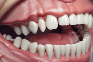
Enamel Structure
- Enamel is the hardest calcified tissue, covering the crown of teeth, with 96% inorganic components (inorganic hydroxyapatite crystals).
- Enamel consists of Enamel Rods (structural units running from the dentinoenamel junction):
- Enamel rods segments are divided by striations.
- A segment is 4 microns in length.
- The Head/Body/Tail are parts of the enamel rod that reflect incremental daily deposition
- Enamel structure is studied through ground sections, as decalcified sections lose the enamel due to its high mineral content.
Enamel Lines and Structures
- Striae of Retzius: Incremental lines representing successive deposition of enamel. Appear as brownish bands, especially visible on cusps and incisal regions, but also as oblique lines in cervical areas. Reflect variation in enamel structure and mineralisation
- Neonatal Line: A prominent incremental line that separates prenatal and postnatal enamel. Develops when major changes in the environment occur during tooth formation. Clear in most deciduous teeth and the first permanent molars.
- Enamel Lamellae: Leaf-like structures extending from the enamel surface to dentin. Hypocalcified and formed in planes of tension, and are further categorized into 3 types.
- Enamel Tufts: Ribbon-like structures extending from DEJ in various directions as one-third to one-fifth of the enamel thickness. Greater organic components.
- Enamel Spindles: Odontoblastic processes crossing the DEJ. Shaped like spindles and approximately 10µm in length.
- Hunter-Schreger Band: Alternate light and dark bands of enamel. Caused by abrupt changes in the direction of enamel rods. The arrangement of enamel rods in the cusp area are more irregular.
- Gnarled Enamel:Wavy course as it extends from the dentinoenamel junction towards the outer surface. Irregular arrangement and intertwining creating 'gnarled' appearance, especially in cusp/incisal areas.
Dentin and Dentinogenesis
- Dentin formation (dentinogenesis) starts during the late bell stage of tooth development. This process occurs in two phases
- Collagen matrix formation: Odontoblasts deposit collagen and other components of extra-cellular matrix.
- Mineralization (calcification): Hydroxyapatite deposits on the collagen fibrils.
Dentin Types
- Primary Dentin: Formed before tooth root formation and in the eruption period
- Secondary Dentin: Formed after the completion of the root formation and eruption of the tooth. Its deposition slower in comparison with primary dentin
- Tertiary Dentin: Reactionary or reparative Dentin. Reactionary is deposited by pre-existing odontoblasts. Reparative is deposited by newly differentiated odontoblasts.
- Types of dentin (detailed): predentin, mantle dentin, circumpulpal dentin, peritubular dentin, intertubular dentin, interglobular dentin, and Tomes granular layer
Dentin Features
- Innervation: Dentin is highly sensitive.
- Incremental lines: Daily matrix deposition.
- Contour lines: Disturbances in matrix & mineralization processes.
- Neonatal line: Marks prenatal and postnatal dentin separation.
Secondary Curvatures
- With age, functional requirements lead to changes in the dentin:
- Secondary dentin formation
- Sclerotic dentin: Dentin with calcified tubules due to injury/aging
- Reparative dentin (Tertiary) formation
- Dead tracts: Loss of odontoblasts, leaving air in dentinal tubules.
Dentin Structure
- Dentin is a hard tissue present in the crown and root of teeth. Its covered by enamel in the crown and cementum in the root.
- General characteristics such as yellow color, slight elasticity, permeability (traversed by tubules), and its composition
- Structures of dentin:
- Dentinal tubules: Span from enamel/cementum to pulp.
- Odontoblasts: Cells that secrete dentin matrix.
- Odontoblastic processes: extend in tubules.
Function of Odontoblasts process
- Secrete hydroxyapatite crystals for mineralization of dentin matrix.
- Secrete tubular dentin.
- Maintain dentinal tubule fluid.
- Produce sclerotic dentin.
- Maintain dentin sensitivity.
Studying That Suits You
Use AI to generate personalized quizzes and flashcards to suit your learning preferences.