Podcast
Questions and Answers
What is the primary process involved in gas exchange?
What is the primary process involved in gas exchange?
- Chemical reaction of gases
- Forced movement of gases
- Active transport of gases
- Passive movement of gases across membranes (correct)
What is the process of capillary gas exchange in body tissues known as?
What is the process of capillary gas exchange in body tissues known as?
- Pulmonary diffusion
- External respiration
- Alveolar ventilation
- Internal respiration (correct)
Which of the following is a key component of gas exchange in the alveoli?
Which of the following is a key component of gas exchange in the alveoli?
- Concentration of nitrogen
- Thickness of the capillary walls
- Partial pressure gradients of gases (correct)
- Active transport of oxygen
What is external respiration?
What is external respiration?
What are the two main factors which drive gas exchange?
What are the two main factors which drive gas exchange?
In the respiratory epithelium, what is the function of goblet cells?
In the respiratory epithelium, what is the function of goblet cells?
The upper respiratory tract is mostly lined with which type of epithelium?
The upper respiratory tract is mostly lined with which type of epithelium?
The nasal vestibule is lined with what kind of epithelium?
The nasal vestibule is lined with what kind of epithelium?
What is the primary function of the respiratory epithelium?
What is the primary function of the respiratory epithelium?
Which cells are primarily responsible for the mucociliary clearance mechanism
Which cells are primarily responsible for the mucociliary clearance mechanism
What is the total percentage of carbon dioxide transported by the blood as a bicarbonate ion?
What is the total percentage of carbon dioxide transported by the blood as a bicarbonate ion?
Carbonic anhydrase is vital to which process?
Carbonic anhydrase is vital to which process?
How much carbon dioxide is transported by the blood dissolved in the plasma?
How much carbon dioxide is transported by the blood dissolved in the plasma?
How many molecules of carbon dioxide can each hemoglobin bind?
How many molecules of carbon dioxide can each hemoglobin bind?
What happens to hemoglobin's affinity for oxygen when carbon dioxide binds to it?
What happens to hemoglobin's affinity for oxygen when carbon dioxide binds to it?
What is the act of air moving in and out of the lungs called?
What is the act of air moving in and out of the lungs called?
During the breathing cycle, what is the brief pause between inspiration and expiration called?
During the breathing cycle, what is the brief pause between inspiration and expiration called?
What determines the direction of air flow during breathing?
What determines the direction of air flow during breathing?
What happens to the intrapleural pressure compared to the alveolar pressure?
What happens to the intrapleural pressure compared to the alveolar pressure?
What is the volume of air that fills the alveoli with an inspiration plus the volume of air that fills the airways?
What is the volume of air that fills the alveoli with an inspiration plus the volume of air that fills the airways?
What structure initiates respiration?
What structure initiates respiration?
Which respiratory group facilitates forced expiration?
Which respiratory group facilitates forced expiration?
What does the pneumotaxic center allow to happen?
What does the pneumotaxic center allow to happen?
Under what conditions is the apneustic center activated, according to the text?
Under what conditions is the apneustic center activated, according to the text?
Which of the following do chemoreceptors monitor?
Which of the following do chemoreceptors monitor?
Intercostal nerves innervate which muscles?
Intercostal nerves innervate which muscles?
During quiet breathing, which muscle increases the length of the thoracic cavity?
During quiet breathing, which muscle increases the length of the thoracic cavity?
What causes air to move into the alveoli during inspiration?
What causes air to move into the alveoli during inspiration?
What happens when the pressure in the alveoli equals the pressure in the atmosphere?
What happens when the pressure in the alveoli equals the pressure in the atmosphere?
What is the effect of decreased diameter of arterioles on pulmonary blood flow?
What is the effect of decreased diameter of arterioles on pulmonary blood flow?
What is the effect on extra alveolar resistance during maximum inspiration?
What is the effect on extra alveolar resistance during maximum inspiration?
What response is triggered by decreased partial pressure of oxygen in the alveoli?
What response is triggered by decreased partial pressure of oxygen in the alveoli?
Where does blood get delivered to in the pulmonary circulation?
Where does blood get delivered to in the pulmonary circulation?
What does the pulmonary blood flow equal?
What does the pulmonary blood flow equal?
What happens to resistance with increased diameter of the arterioles?
What happens to resistance with increased diameter of the arterioles?
What is the approximate value of the tidal volume for adults during normal, quiet breathing?
What is the approximate value of the tidal volume for adults during normal, quiet breathing?
What is the expiratory reserve volume?
What is the expiratory reserve volume?
The trachea consists of how many tracheal cartilages?
The trachea consists of how many tracheal cartilages?
What forms the posterior wall of the trachea?
What forms the posterior wall of the trachea?
What kind of tissue binds the trachea to the adjacent structures?
What kind of tissue binds the trachea to the adjacent structures?
What replaces the cartilage rings in the bronchi?
What replaces the cartilage rings in the bronchi?
Which of the options is not a layer found in the bronchus?
Which of the options is not a layer found in the bronchus?
The bronchioles are characterized by the presence of what kind of epithelium?
The bronchioles are characterized by the presence of what kind of epithelium?
Which substance lines the alveoli walls, reduces the surface tension, and participates in the clearance of foreign materials?
Which substance lines the alveoli walls, reduces the surface tension, and participates in the clearance of foreign materials?
Flashcards
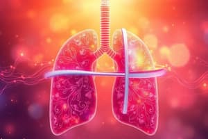
Gas exchange
Gas exchange
Passive movement of gases across membranes.
External respiration
External respiration
Flow of air into the lungs and transfer of oxygen and carbon dioxide through diffusion.
Internal respiration
Internal respiration
Capillary gas exchange in body tissues.
Components of alveolar gas exchange
Components of alveolar gas exchange
Signup and view all the flashcards
Partial pressure gradients
Partial pressure gradients
Signup and view all the flashcards
PA
PA
Signup and view all the flashcards
Pa
Pa
Signup and view all the flashcards
Movement of Oxygen
Movement of Oxygen
Signup and view all the flashcards
Movement of Carbon Dioxide
Movement of Carbon Dioxide
Signup and view all the flashcards
Ventilation vs. Perfusion
Ventilation vs. Perfusion
Signup and view all the flashcards
Gas Exchange in Liquid (Blood)
Gas Exchange in Liquid (Blood)
Signup and view all the flashcards
High PO2, low PCO2
High PO2, low PCO2
Signup and view all the flashcards
Low PO2, high PCO2
Low PO2, high PCO2
Signup and view all the flashcards
External Respiration Summary
External Respiration Summary
Signup and view all the flashcards
Exchange of O2 and CO2
Exchange of O2 and CO2
Signup and view all the flashcards
Oropharynx epithelium
Oropharynx epithelium
Signup and view all the flashcards
Sinuses epithelium
Sinuses epithelium
Signup and view all the flashcards
Epiglottis's lingual vs laryngeal surface
Epiglottis's lingual vs laryngeal surface
Signup and view all the flashcards
True vocal fold epithelium
True vocal fold epithelium
Signup and view all the flashcards
Hyaline cartilage
Hyaline cartilage
Signup and view all the flashcards
CO2 turns into a bicarbonate ion
CO2 turns into a bicarbonate ion
Signup and view all the flashcards
CO2 dissolved in plasma
CO2 dissolved in plasma
Signup and view all the flashcards
CO2 binds to hemoglobin
CO2 binds to hemoglobin
Signup and view all the flashcards
Hemoglobin binding CO2 decreases O2 affinity
Hemoglobin binding CO2 decreases O2 affinity
Signup and view all the flashcards
Ventilation
Ventilation
Signup and view all the flashcards
Inspiration
Inspiration
Signup and view all the flashcards
Expiration
Expiration
Signup and view all the flashcards
Rest period
Rest period
Signup and view all the flashcards
Direction of air flow
Direction of air flow
Signup and view all the flashcards
Tidal volume
Tidal volume
Signup and view all the flashcards
Pulmonary blood flow
Pulmonary blood flow
Signup and view all the flashcards
Alveolar vessels
Alveolar vessels
Signup and view all the flashcards
Vasculature
Vasculature
Signup and view all the flashcards
Histology of the Bronchi
Histology of the Bronchi
Signup and view all the flashcards
Trachealis Muscle
Trachealis Muscle
Signup and view all the flashcards
Histology of the Bronchioles
Histology of the Bronchioles
Signup and view all the flashcards
Terminal Bronchioles
Terminal Bronchioles
Signup and view all the flashcards
Respiratory bronchioles
Respiratory bronchioles
Signup and view all the flashcards
Alveoli
Alveoli
Signup and view all the flashcards
Study Notes
- Gas exchange in lung and body tissues is driven by partial pressures and matching perfusion with ventilation.
Definitions
- Gas exchange involves passive movement of gasses across membranes.
- External respiration is the flow of air into the lungs and the transfer of O2 and CO2 through diffusion.
- Internal respiration is capillary gas exchange in body tissues.
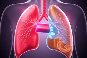
Mechanisms of Alveolar Gas Exchange
- The three components include the surface area of the alveolo-capillary membrane.
- Gas exchange also depends on partial pressure gradients of the gasses
- Matching ventilation and perfusion are also key
Gas Exchange Across Alveolo-capillary Membrane
- Partial pressure gradients are the difference between partial pressure of gas in the alveolar sac and the blood.
- This gradient is the driving force for gas diffusion across the membrane.
- It is also called the Partial Pressure Gradient, not the concentration difference.
- PA represents partial pressure of gas in the alveolar sac.
- Pa is partial pressure of a gas in the blood.
Movement of Oxygen
- Oxygen diffuses from high to low partial pressure areas, moving from alveoli into blood.
- PAO2 > PaO2, the pressure gradient is 60mmHg.
Movement of Carbon Dioxide
- Carbon dioxide diffuses from high to low partial pressure, moving from capillary into alveoli.
- PACO2 < PaCO2, the pressure gradient is 5mmHg.
Movement of a Gas Between Air and a Liquid (Blood)
- The exchange of CO2 from blood into alveoli during inspiration is facilitated this way.
- The amount of gas dissolving in a liquid is directly proportional to its partial pressure in the air above.
- High partial pressure results in a high amount of dissolved gasses by volume.
- Gases can be forced to dissolve into a liquid if enough pressure is applied
- Gases can leave a liquid if external pressure lowers.
Inspiration
- Alveolar pressure decreases causing CO2 dissolved in blood to leave and return to a gaseous state.
Matching of Ventilation and Perfusion
- Ventilation refers to gas reaching alveoli.
- Perfusion refers to blood flow in pulmonary capillaries servicing alveoli.
Factors Affecting Airway and Blood Vessel Diameters
- PACO2 controls the diameter of bronchioles.
- PAO2 controls the diameter of blood capillaries.
Alveoli Conditions
- High PAO2 and low PACO2 leads to bronchioles constricting and pulmonary capillaries dilating.
- Low PO2 and high PCO2 leads to bronchioles dilating and pulmonary capillaries constricting.
External Respiration Summary
- Mixed venous blood returns from tissues to the right heart and is then pumped to the pulmonary artery before reaching pulmonary capillaries.
- The PCO2 level in this blood averages 45 mm Hg and PO2 40 mm Hg, reflecting tissue metabolic activity.
- This is because cellular activity produces CO2 and consumes O2, leading to PCO2 > PO2.
- O2 and CO2 exchange occurs between alveolar air and capillary blood.
- Blood leaving pulmonary capillaries normally has the same PO2 and PCO2 as alveolar air.
- PaO2 is 100 mm Hg and PaCO2 is 40 mm Hg
- The oxygenated blood returns to the left heart and is pumped into the aorta for systemic circulation.
Internal Respiration
- A partial pressure gradient exists between blood and tissues.
- Oxygen partial pressure is low in tissues (40 mm Hg) and high in the blood (100 mm Hg).
- Oxygen diffuses out of the blood due to this gradient.
- Carbon dioxide diffuses into the blood because its partial pressure is lower in the blood.
Nasal Cavity: Histology
- Most of the upper respiratory tract is lined with pseudostratified ciliated columnar epithelium, also known as respiratory epithelium.
- Exceptions include parts of the pharynx and larynx.
- All cells contact the basement membrane.
- Epithelium appears multi-layered due to non-alignment of cell nuclei, hence the term pseudostratified.
- There are five cell types, but only three will be focussed on, and they are: ciliated columnar, goblet, and basal cells.
- Ciliated columnar cells are the most abundant, extending the epithelium.
- Cilia sweep the surface to remove inhaled particles and protect the lungs.
- Goblet cells are columnar epithelial cells shaped like wine goblets which contain and secrete mucus glycoproteins.
- Mucus forms a protective layer, maintains moisture, and traps particles and pathogens.
- Located in proximal airways and decreasing in numbers distally.
- Basal cells are small, cubodial cells near the basal lamina.
- Their apices does not extend to the lumen.
- These function as stem cells.
Function of the Respiratory Epithelium
- It works to moisten and protect the airways, acting as a physical barrier to pathogens and their removal.
- The ciliated cells are key in mucociliary clearance. Each epithelial cell has ~200 cilia beating 10-20 times per second.
- The beat is targeted towards the pharynx, either upwards from the lower respiratory tract, or downwards from the nasal structures.
Vestibule of the Nasal Cavity
- It is part of the external nose and communicates with the external environment.
- The epithelial lining is stratified squamous epithelium, a continuation of facial skin, containing vibrissae to entrap large particles.
- Sebaceous glands assist with this entrapment.
- Posteriorly, stratified squamous epithelium thins and transitions into the pseudostratified epithelium characteristic of the respiratory region.
- Sebaceous glands are absent there.
- Stratified squamous epithelium consists of several layers where the apical layer and several layers beneath, are squamous.
- Deeper, the cells range from cubodial to columnar.
- Non-keratinized stratified squamous epithelium cells do not have much keratin and are moisturized by mucus from salivary or mucous glands.
- Flattened cells retain nuclei and metabolism.
Respiratory Region of the Nasal Cavity
- This area constitutes most of the nasal cavities, and has a lining of respiratory epithelium, that being pseudostratified ciliated columnar epithelium with goblet cells.
- The lamina propria of the respiratory mucosa has many blood vessels.
- Vessel arrangement allows warming of inhaled air.
- These vessels can become engorged and leaky during allergic reactions/viral infections.
- This may cause fluid distension, resulting in swelling and breathing difficulties.
- It also contains mucous glands.
- The glands have ducts that open onto the epithelial surface, supplementing goblet cells.
- The area has conchae (turbinates) to increase the efficiency in warming inspired air by increasing surface area.
- Particles in the air stream adhere to mucus-covered walls.
- Particles trapped here are transported to the pharynx via ciliary movements and are swallowed, or forcefully expelled through sneezing.
Olfactory Region of the Nasal Cavity
- Found on part of the dome of each nasal cavity, lined with specialized olfactory mucosa.
- Human olfactory mucosa is only 10 cm², whereas animals with a good sense of smell can have over 150 cm² such as certain dog species.
- The lamina propria is contiguous with the periosteum of the underlying bone and contains many blood and lymphatic vessels, unmyelinated and myelinated olfactory nerves, and olfactory glands.
- Olfactory epithelium is pseudostratified tall columnar epithelium without goblet cells or motile cilia.
- It is composed of olfactory receptor cells, supporting cells, and basal cells.
- Olfactory receptor cells are neurons spanning the epithelium and entering the central nervous system.
- Supporting cells are columnar cells for mechanical and metabolic support to the olfactory receptor cells.
- They synthesize and secrete odorant-binding proteins.
- Basal cells are stem cells that differentiate into new olfactory receptor and supporting cells.
Transitional Area
- Transition occurs between both the olfactory and respiratory epithelium is abrupt.
Paranasal Sinuses: Histology
- The mucosal lining is thin, ciliated, pseudostratified columnar epithelium with numerous goblet cells.
- The lamina propria is thin and continuous with the periosteum, containing few small glands.
- Mucus produced is swept into the nasal cavities via ciliary movements.
Pharynx: Histology
- The nasopharynx epithelium is continuous with the nasal cavity.
- The oropharynx has non-keratinized stratified squamous epithelium.
- This durable type is better suited for friction associated with swallowing food.
- Lymphatic aggregates in the mucosa act as first contact points for the immune system to check entering particles.
Larynx: Histology
- The epiglottis has both lingual and laryngeal surfaces on the anterior and posterior sides respectively.
Epiglottis
- Elastic cartilage forms the framework, allowing the structure to snap back into shape.
- This provides strength, elasticity, and more flexibility.
- The lingual mucosa has stratified squamous nonkeratinized epithelium.
- The lamina propria merges with the perichondrium of the elastic cartilage.
- The lingual mucosa covers the apex and half of the laryngeal surface.
- Towards the base of the laryngeal surface, it changes to pseudostratified ciliated columnar epithelium.
- Taste buds and lymphatic nodules may be present in both the lingual and laryngeal epithelium.
Larynx
- The region from epiglottis to true vocal fold is lined by respiratory epithelium, or pseudostratified ciliated columnar epithelium with goblet cells.
- The lamina propria has mixed seromucous glands which empty onto the epithelial surface.
- The true vocal fold mucosa has nonkeratinized stratified squamous epithelium and a thin, dense lamina propria without glands, lymphatic tissue, or blood vessels.
- Inferior to the true vocal fold, the epithelium switches back to respiratory epithelium (pseudostratified ciliated columnar epithelium with goblet cells). The lamina propria has seromucous glands. The hyaline cricoid cartilage is at the bottom.
Thyroid Cartilage and Cricoid Cartilage
- both cartilages are composed of hyaline cartilage, containing sparse chondrocytes surrounded by extracellular matrix.
- A high water content gives the matrix a glossy appearance, and gives the cartilage a flexible yet firm support.
Carbon Dioxide Transport
- CO2 is transported through three mechanisms: dissolved in plasma, binds to hemoglobin and converted into a bicarbonate ion.
Dissolved CO2
- A small amount of CO2 totaling 5% of the transport.
CO2 Binding to Hemoglobin
- CO2 binds to amino acids on globin chains, making up 3% of its transport.
- Each hemoglobin can bind four molecules of CO2.
- Carboaminohemoglobin occurs when hemoglobin binds to carbon dioxide and decreases its affinity for oxygen.
- This leads to O2 becoming unbound and getting dropped off in tissues full of CO2 resulting in a shift to the right on the oxygen-hemoglobin dissociation curve.
CO2 Conversion
- Around 90% of CO2 is turned into bicarbonate ions (HCO3-).
- Carbonic anhydrase in RBCs produces HCO3- and H+.
- As bicarbonate ion levels build up in red blood cells, the ions move into the plasma.
- Cl- then comes into the RBC going into the plasma (chloride shift)
Bicarbonates
- Bicarbonates help maintain physiologic H+ levels in the blood.
- This follows the LeChatelier principle.
- Low hydrogen ion concentration shifts the equation to the right, generating more bicarbonate and hydrogen ions.
- High hydrogen ion concentration causes bicarbonate to bind to form carbonic acid and separate, forming water and carbon dioxide.
- The hydrogen ion concentration does not shift too quickly in either direction, which means that pH levels remain stable.
CO2 in Lungs
- CO2 leaves the blood, so more O2 diffuses and binds to hemoglobin in RBCs.
- Hemoglobin's affinity for CO2 decreases which causes CO2 and H+ to become unbound.
- A rise in H+ encourages more HCO3- to bind with it, with HCO3 re-entering the red blood cell from the plasma.
- This increases PCO2 and its partial pressure gradient, resulting in carbon dioxide entering the alveoli.
Breathing Mechanics
Components of Breathing
- Ventilation is how air moves in and out of the lungs.
- Inspiration is the process of air flowing into the lungs.
- Expiration is the process of air leaving the lungs.
- The rest period is a brief pause between inspiration and expiration.
Pressures
- Airflow direction is determined by the atmospheric pressure and alveolar pressure difference.
- Atmospheric pressure is the pressure of air in the environment
- Alveolar pressure is the pressure of air inside the alveoli
- Intrapleural pressure (intrathoracic pressure) is the fluid pressure inside the pleural cavity around the lungs. It is usually negative compared to alveolar or atmospheric pressures.
- Transmural pressure is alveolar pressure minus intrapleural pressure.
- When transmural pressure is positive, airways remain open.
- Lung and chest walls act as opposing forces, with lungs tending to collapse and chest walls expand during rest.
- Tidal volume measures the volume of air that fills the alveoli with inspiration plus the volume of air that fills the airways.
Respiratory Centers
- Groups of neurons control ventilation with the medulla oblongata.
- The dorsal respiratory group initiates respiration and sets the breathing rhythm.
- The ventral respiratory group facilitates forced expiration.
- These groups send inhibitory impulses to the apneustic center in the pons.
- Decreasing the duration of inspiration can allow leaves more breathing time for the body.
- The pontine respiratory group consist of
- Pneumotaxic center which allows expiration to happen.
- When activated, it limits the DRG activation which decreases action potentials of the phrenic nerve and stops inspiration.
- The Apneustic center
- It activates when the body requires more oxygen that stimulates neurons in the apneustic to excite the dorsal respiratory group.
- This prolongs action potentials in the phrenic nerve to prolong the contraction of the diaphragm for prolonged inspiration.
Chemoreceptors
- Chemoreceptors monitor CO2 and O2 concentration in the blood.
- Abnormal concentrations cause signals to be sent to the respiratory center neurons.
- Normal values include PaO2 at 100 mmHg, PaCO2 at 40 mmHg, and arterial pH at 7.4.
Muscles
- The diaphragm increases the length of thoracic cavity Quiet breathing relies on
- External intercostal muscles Pull ribs up and out to increase the thorax width
Accessory
- Sternocleidomastoid
- Scalene muscles
- These contract during vigorous inspiration to move ribs up and out.
Nerves
- The Phrenic nerve innervates the diaphragm.
- The Intercostal nerves provide innervation to the external intercostal muscles.
Breathing Cycle Rest Phase.
- Diaphragm is at rest and alveolar pressure equals atmospheric pressure, meaning air is not moving.
- Transmural pressure is at +5cm, keeping airways open
Inspiration
- PaCO2 levels increase.
- If detected by chemoreceptors, the dorsal respiratory group responds via phrenic nerve, activating the diaphragm and intercostal nerves.
- This stimulates external intercostal muscles, causing contraction and increasing thorax volume.
- Lung volume increases lead to alveolar pressure decreasing
- Decreasing it below atmospheric pressure creates a pressure gradient, drawing air in.
- Flow stops when Pressure in the Alveoli = Pressure in the atmosphere.
Expiration
- Diaphragm Muscles relax causing air to leaves the lungs as they return to their normal size
- This leads to alveolar pressure increasing, as it exceeds atmospheric pressure again creating new pressure gradient and allowing the air out the lungs.
- Eventually, all volumes and pressures return to initial rest phase.
Pulmonary Circulation
- Involves blood flow starting from the right ventricle which pumps out to the pulmonary trunk then arrives at the pulmonary arteries, and arterioles. Capillaries then surround alveoli.
- Pulmonary capillaries drains into small veins and eventually form the two pulmonary veins exiting each lung.
- This delivers oxygen-rich blood into the left atrium.
- Pulmonary blood flow (Q) is the volume of blood pumped out the ventricle during a time interval.
- Usually one minute
- The Cardiac output of the right ventricle is also relevant
- mL per heartbeat x heart rate (beats per minute)
- Directly proportional to the difference in pressure: pulmonary artery and left atrium = ΔΡ
- Inversely proportional to the resistance of the pulmonary vasculature (R)
- Systemic Blood Pressure>Pulmonary Blood Pressures in Circulation
Resistance of vasculature
- Changes in resistance are controlled by the diameter of the arterioles.
- Decreased diameter of arterioles increases of vascular resistance leading to decreased blood flow.
- Increased diameter of arterioles decreases in vascular resistance thereby increasing blood flow.
Total Resistance of Pulmonary Vasculature
- Depends on alveolar vessels, the extra-alveolar vessels and the air pressure.
Types of Vessels
- Alveolar vessels are tiny capillaries surrounding the alveoli.
- The extra-alveolar vessels are arterioles that are located further away.
Air Pressure
- Alveolar air pressure being in high volumes will close alveolar blood vessels.
- Low volumes of air will let blood vessel open.
- Maximum inspiration involves the extra-alveolar reducing vessels due to the lung outward expansion.
- Air pressure in the pleural will be lower to reduce volume of air.
Summary
- Total pulmonary Resistance = Sum of Alveolar Vessel with Extra-Alveolar Resistance
- Highest total pulmonary resistance involves
- Maximum inspiration = alveolar resistance is high.
- Low levels of Air in Total Pulmonary Resistance points before expiration or inspiration occurs
- Maximum Expiration = Extra-Alveolar Resistance is high.
- Resistance controlled by smooth which regulates vasodilation and constriction around arterioles and around the vasculature.
Pulmonary Arteriole Constriction and Dilation
- Vasoconstriction and vasodilation are controlled by the smooth muscle around the arterioles.
Vasoconstriction
- Increased in resistance to airflow which lowers partial pressure due to the oxygen volume around the alveoli.
- Called Hypoxic Vasoconstriction.
- Diseased or damaged air with pneumonia can activate this vasoconstriction and close off the areas of airflow where it is not able to produce oxygen.
- Blood can be oxygenated efficiently if this happens
- Hypoxic happens, vessels throughout whole lung close and the right side of the heart increase pressure due to no proper oxygenation in circulation.
Vasodilation
- Decreased vascular resistance.
Vasoactive
- Created with cells that increase vascularization.
- Can bind to and affect smooth muscle cells.
Factors that Influence Vasoconstriction
- Made with a binding of cells, it creates an abundance.
- Thromboxane increases vascularization
- Produced by leukocytes in the lung tissues.
- Prostaglandin increases dilation.
- Produced with Lung of endothelial.
- Nitric oxide helps smooth muscle cell vasodilate.
Lung Volumes (Tidal Volume, Inspiratory/Expiratory ReserveVolume, Residual Volume and Lung Capacities (Functional Residual Capacity, Inspiratory Capacity, Vital Capacity, Total Lung Capacity)
- All volumes measure to help quantify airflow in relation air going in and out during breaths.
LUNG VOLUMES
Tidal Volume
- Represents how the air moves whether inhaling or exhaling when take breaths.
- During rest or "quiet" regular breathing cycle, it takes about 500 mL to breathe.
Inspiratory volume
- Volume of air is more than what can inhaled above the level of tidal volume.
- Usually only happens with peak max effort of heavy breathing where volumes reach higher numbers.
- This provides that max level backup volume, a safety for respiration, where there is not usually the need to use this state unless pushed to one's limit.
- Average in volume is 3 liters
Expiratory volume
- Volume of air that gets from what can be exhaled which stays below that volume.
- Average in at with volumes equal to 1.2 liters.
Residual volume
- Volume that has air lasting maximum peak efforts.
- Average around volume with total volumes equal 1.2 liters to air remains in during expiration cycles maximum effort, lung would be empty with amount.
Lung Capacities
- Capacities of these volumes helps helps measure volume amount where lungs functions and operate
- Functional Residual, - Expiratory reserve volume with residual volume average near 2.4L of air is
- Measured with at volume with at equal to 3.5 liters, tidal volume, tidal measures average liters with 3.5 in tidal and in litres
- "Total liters" also equals tidal volume, litres the amount there, average measure total as amount there
- Volume of equal to .9 l with measure with is the total the for hold much litres. Is equal to and the hold
Studying That Suits You
Use AI to generate personalized quizzes and flashcards to suit your learning preferences.