Podcast
Questions and Answers
After delivering oxygen and nutrients, what does blood primarily collect for transport back to the lungs?
After delivering oxygen and nutrients, what does blood primarily collect for transport back to the lungs?
- Water
- Hormones
- Carbon dioxide (correct)
- Electrolytes
How does blood regulate body temperature?
How does blood regulate body temperature?
- Maintaining a constant blood viscosity.
- Decreasing blood flow to the skin to retain heat.
- Increasing blood flow to the skin to dissipate heat. (correct)
- Regulating electrolyte balance.
Besides plasma and red blood cells, what is the other component of whole blood?
Besides plasma and red blood cells, what is the other component of whole blood?
- Serum
- Interstitial fluid
- Lymph
- Buffy coat (correct)
What is the approximate percentage of plasma in whole blood?
What is the approximate percentage of plasma in whole blood?
Which components are found within the buffy coat layer of a centrifuged blood sample?
Which components are found within the buffy coat layer of a centrifuged blood sample?
How does the hematocrit level typically differ between adult men and women?
How does the hematocrit level typically differ between adult men and women?
Albumin is a protein found in blood plasma. What is its primary role?
Albumin is a protein found in blood plasma. What is its primary role?
Which type of globulins primarily functions in immunity?
Which type of globulins primarily functions in immunity?
What happens to the heme that is leftover after red blood cell breakdown?
What happens to the heme that is leftover after red blood cell breakdown?
Which of the following is a primary characteristic of anemia?
Which of the following is a primary characteristic of anemia?
A patient is diagnosed with hemorrhagic anemia. What is the underlying cause of this condition?
A patient is diagnosed with hemorrhagic anemia. What is the underlying cause of this condition?
Iron deficiency anemia is often a secondary result of what other type of anemia?
Iron deficiency anemia is often a secondary result of what other type of anemia?
What is the primary cause of pernicious anemia?
What is the primary cause of pernicious anemia?
How does renal anemia develop in patients with kidney damage or failure?
How does renal anemia develop in patients with kidney damage or failure?
What underlying process defines hemolytic anemia?
What underlying process defines hemolytic anemia?
In sickle cell anemia, what specific molecular change leads to the altered shape of red blood cells?
In sickle cell anemia, what specific molecular change leads to the altered shape of red blood cells?
What would occur if the chordae tendineae and papillary muscles failed to function properly during ventricular contraction?
What would occur if the chordae tendineae and papillary muscles failed to function properly during ventricular contraction?
How do the semilunar valves prevent backflow of blood into the ventricles?
How do the semilunar valves prevent backflow of blood into the ventricles?
During ventricular contraction, why do the semilunar valves open?
During ventricular contraction, why do the semilunar valves open?
Which of the following statements accurately describes the structure of the semilunar valves?
Which of the following statements accurately describes the structure of the semilunar valves?
What is the primary reason the heart needs its own coronary circulation despite being filled with blood?
What is the primary reason the heart needs its own coronary circulation despite being filled with blood?
Which area of the heart does the anterior interventricular artery supply with blood?
Which area of the heart does the anterior interventricular artery supply with blood?
A patient is diagnosed with a blockage in the left coronary artery (LCA) before it bifurcates. Which of the following is the MOST likely consequence?
A patient is diagnosed with a blockage in the left coronary artery (LCA) before it bifurcates. Which of the following is the MOST likely consequence?
What is the primary function of the coronary circulation?
What is the primary function of the coronary circulation?
Which characteristic of erythrocytes is most directly related to their function of efficient gas exchange?
Which characteristic of erythrocytes is most directly related to their function of efficient gas exchange?
During erythropoiesis, at what stage does the cell eject its nucleus and organelles, leading to its biconcave shape?
During erythropoiesis, at what stage does the cell eject its nucleus and organelles, leading to its biconcave shape?
If a patient has a condition that reduces the production of erythrocytes, what hormone or nutrient deficiencies might be contributing factors?
If a patient has a condition that reduces the production of erythrocytes, what hormone or nutrient deficiencies might be contributing factors?
How does the structure of hemoglobin facilitate its role in oxygen transport?
How does the structure of hemoglobin facilitate its role in oxygen transport?
What happens to the components of erythrocytes when they are broken down in the spleen?
What happens to the components of erythrocytes when they are broken down in the spleen?
A researcher is studying blood samples and observes cells without nuclei. Which type of formed element are they most likely observing?
A researcher is studying blood samples and observes cells without nuclei. Which type of formed element are they most likely observing?
What is the key distinction between blood plasma and blood serum?
What is the key distinction between blood plasma and blood serum?
If red bone marrow activity is severely compromised, which of the following would be the most immediate and concerning consequence?
If red bone marrow activity is severely compromised, which of the following would be the most immediate and concerning consequence?
During resting conditions, the parasympathetic nervous system (PNS) reduces heart rate through which mechanism?
During resting conditions, the parasympathetic nervous system (PNS) reduces heart rate through which mechanism?
How do elevated levels of potassium ions affect heart function?
How do elevated levels of potassium ions affect heart function?
What is the primary mechanism by which exposure to cold temperatures directly decreases heart rate?
What is the primary mechanism by which exposure to cold temperatures directly decreases heart rate?
Which of the following is a characteristic of tachycardia?
Which of the following is a characteristic of tachycardia?
Which factor is most likely to result in bradycardia?
Which factor is most likely to result in bradycardia?
How does coronary atherosclerosis contribute to homeostatic imbalance of cardiac output?
How does coronary atherosclerosis contribute to homeostatic imbalance of cardiac output?
What is the primary effect of persistent high blood pressure on the myocardium?
What is the primary effect of persistent high blood pressure on the myocardium?
If a patient has chronically elevated afterload due to hypertension, what happens to the end systolic volume (ESV)?
If a patient has chronically elevated afterload due to hypertension, what happens to the end systolic volume (ESV)?
Which of the following accurately describes the role of the endomysium within cardiac muscle tissue?
Which of the following accurately describes the role of the endomysium within cardiac muscle tissue?
How do desmosomes in intercalated discs contribute to cardiac muscle function?
How do desmosomes in intercalated discs contribute to cardiac muscle function?
What is the functional significance of gap junctions in cardiac muscle?
What is the functional significance of gap junctions in cardiac muscle?
Why is the resistance to fatigue important in cardiac cells?
Why is the resistance to fatigue important in cardiac cells?
How does the presence of gap junctions in cardiac muscle, compared to their absence in skeletal muscle, affect the overall function of the respective tissues?
How does the presence of gap junctions in cardiac muscle, compared to their absence in skeletal muscle, affect the overall function of the respective tissues?
If the gap junctions in cardiac muscle were compromised, what would be the most likely consequence?
If the gap junctions in cardiac muscle were compromised, what would be the most likely consequence?
Cardiac muscle utilizes sarcomeres. Sarcomeres have Z discs, A bands, and I bands that reflect the arrangement of the thick (myosin) and thin (actin) filaments composing them. How does this arrangement specifically contribute to the function of cardiac muscle?
Cardiac muscle utilizes sarcomeres. Sarcomeres have Z discs, A bands, and I bands that reflect the arrangement of the thick (myosin) and thin (actin) filaments composing them. How does this arrangement specifically contribute to the function of cardiac muscle?
Which of the following is a key functional difference between cardiac and skeletal muscle regarding their stimulation and contraction mechanisms?
Which of the following is a key functional difference between cardiac and skeletal muscle regarding their stimulation and contraction mechanisms?
Flashcards
Blood's primary transport functions
Blood's primary transport functions
Transports oxygen and picks up carbon dioxide. Transports nutrients and hormones.
Blood's regulation functions
Blood's regulation functions
Regulates body temperature, pH balance, and fluid volume in the circulatory system.
Blood's protective functions
Blood's protective functions
Prevents blood loss and protects against infection.
Major blood components
Major blood components
Signup and view all the flashcards
Physical characteristics of blood
Physical characteristics of blood
Signup and view all the flashcards
Composition of whole blood
Composition of whole blood
Signup and view all the flashcards
Buffy coat composition
Buffy coat composition
Signup and view all the flashcards
Hematocrit
Hematocrit
Signup and view all the flashcards
Bilirubin
Bilirubin
Signup and view all the flashcards
Anemia
Anemia
Signup and view all the flashcards
Hemorrhagic Anemia
Hemorrhagic Anemia
Signup and view all the flashcards
Iron Deficiency Anemia
Iron Deficiency Anemia
Signup and view all the flashcards
Pernicious Anemia
Pernicious Anemia
Signup and view all the flashcards
Renal Anemia
Renal Anemia
Signup and view all the flashcards
Hemolytic Anemia
Hemolytic Anemia
Signup and view all the flashcards
Thalassemia
Thalassemia
Signup and view all the flashcards
Fibrinogen
Fibrinogen
Signup and view all the flashcards
Serum
Serum
Signup and view all the flashcards
Formed Elements
Formed Elements
Signup and view all the flashcards
Erythrocytes Characteristics
Erythrocytes Characteristics
Signup and view all the flashcards
Hemoglobin
Hemoglobin
Signup and view all the flashcards
Erythrocyte Creation
Erythrocyte Creation
Signup and view all the flashcards
Erythropoiesis
Erythropoiesis
Signup and view all the flashcards
Erythrocyte Recycling
Erythrocyte Recycling
Signup and view all the flashcards
Chordae Tendineae
Chordae Tendineae
Signup and view all the flashcards
Papillary Muscles
Papillary Muscles
Signup and view all the flashcards
Semilunar (SL) Valves
Semilunar (SL) Valves
Signup and view all the flashcards
Aortic Valve
Aortic Valve
Signup and view all the flashcards
Pulmonary Valve
Pulmonary Valve
Signup and view all the flashcards
SL Valve Structure
SL Valve Structure
Signup and view all the flashcards
Coronary Circulation
Coronary Circulation
Signup and view all the flashcards
Left Coronary Artery (LCA)
Left Coronary Artery (LCA)
Signup and view all the flashcards
SNS & Exercise
SNS & Exercise
Signup and view all the flashcards
Heat & HR
Heat & HR
Signup and view all the flashcards
PNS Activation
PNS Activation
Signup and view all the flashcards
Vagal Tone
Vagal Tone
Signup and view all the flashcards
High Potassium & HR
High Potassium & HR
Signup and view all the flashcards
Cold & Heart Rate
Cold & Heart Rate
Signup and view all the flashcards
Tachycardia
Tachycardia
Signup and view all the flashcards
Bradycardia
Bradycardia
Signup and view all the flashcards
Endomysium
Endomysium
Signup and view all the flashcards
Intercalated Discs
Intercalated Discs
Signup and view all the flashcards
Desmosomes (in cardiac muscle)
Desmosomes (in cardiac muscle)
Signup and view all the flashcards
Gap Junctions (in cardiac muscle)
Gap Junctions (in cardiac muscle)
Signup and view all the flashcards
Functional Syncytium
Functional Syncytium
Signup and view all the flashcards
Cardiac Muscle: Gap Junctions
Cardiac Muscle: Gap Junctions
Signup and view all the flashcards
Skeletal Muscle: No Gap Junctions
Skeletal Muscle: No Gap Junctions
Signup and view all the flashcards
Why Cardiac Syncytium is Necessary
Why Cardiac Syncytium is Necessary
Signup and view all the flashcards
Study Notes
- Blood transports oxygen, nutrients, and hormones to the body, and removes carbon dioxide.
- Blood regulates body temperature, pH balance, and fluid volume.
- Blood prevents blood loss and infections.
Blood Composition
- The two major components of whole blood are plasma and packed red blood cells.
- Blood is connective tissue with plasma as the matrix.
- Dissolved fibrous proteins in plasma become fibrin strands during clotting.
- Blood is a sticky, opaque fluid with a metallic taste, making up about 8% of body weight.
- Adult men have 5-6 L of blood, while adult women have 4-5 L.
- Whole blood consists of 55% plasma, less than 1% buffy coat, and 45% packed red blood cells.
- Buffy coat contains leukocytes (WBCs) and platelets for immunity and clotting.
- Hematocrit, or blood fraction, is the total volume of blood within a sample, approximately 47% in men and 42% in women.
Plasma Components
- Plasma contains water, electrolytes, proteins, and other substances like nutrients and gases.
- Water and electrolytes maintain osmotic pressure and pH balance.
- Plasma proteins include albumin, globulins, and fibrinogen.
- Albumin maintains osmotic pressure.
- Globulins (alpha, beta, and gamma) transport proteins and contain antibodies.
- Fibrinogen forms fibrin for clotting. Serum is plasma without proteins.
Formed Elements
- Formed elements are cells and cell fragments suspended in plasma, including erythrocytes, leukocytes, and platelets.
- Erythrocytes lack nuclei and organelles, platelets are cell fragments, and leukocytes are complete cells.
- Most formed elements survive only a few days and are replaced by stem cells from red bone marrow.
- Erythrocytes' shape and small size increase their surface area for gas exchange.
Hemoglobin
- Hemoglobin consists of heme (red pigment w/ iron) bound to protein globin.
- Globin contains 4 polypeptide chains (2 alpha, 2 beta), enabling oxygen transport.
Erythrocyte Lifecycle
- Erythrocytes circulate for about 120 days.
- Erythropoiesis takes about 15 days.
- Stem cells in bone marrow become proerythroblasts, then basophilic erythroblasts where ribosomes are synthesized.
- Hemoglobin is synthesized and iron accumulates as the basophilic erythroblast develops into polychromatic then orthochromatic erythroblasts.
- The nucleus is ejected during the process forming a reticulocyte which matures into erythrocyte.
- Erythropoiesis, is controlled by hormones and requires amino acids, iron, and B12 vitamins.
- Damaged erythrocytes are sent to the spleen, where iron is recycled for new erythrocytes.
- Heme becomes bilirubin, which is sent to the liver, and carbon dioxide is exhaled.
Anemia
- Anemia is a compromised oxygen-carrying capacity.
- Hemorrhagic anemia is due to rapid blood loss.
- Iron deficiency anemia is the most common and caused by lack of iron.
- Pernicious anemia is an autoimmune disorder affecting older adults.
- Renal anemia is caused by lack of EPO due to kidney damage.
- Aplastic anemia is where the production of RBCs is stopped.
- Hemolytic anemia is excessive RBC destruction, potentially caused by genetic disorders such as Thalassemia.
- Thalassemia is a genetic disorder causing thin, delicate, hemoglobin deficient RBCs.
- Sickle cell anemia is a genetic disorder causing abnormal hemoglobin, resulting in sickle-shaped RBCs, leads to organ failure etc.
Polycythemia
- Polycythemia is an excess of erythrocytes increases blood volume.
- It increases risk clotting and stroke and can be treated with medication or phlebotomy.
Leukocytes Function
- Leukocytes counter infections and include bacterial, viral, parasitic, toxic, and tumor cells.
- Diapedesis is the action of the white blood cells slipping out of capillary blood vessels.
- Amoeboid motion assists movement to affected areas through tissue spaces.
- Positive chemotaxis pinpoints damage or infections for destruction.
- Neutrophils are most common and fight bacteria. Eosinophils kill parasitic worms, and basophils release histamine.
- Lymphocytes attack via direct cell or antibodies, and monocytes become macrophages.
Leukemia
- Leukemia is abnormal leukocyte overproduction, forming cancerous cells.
- These cells will attack the lymph nodes and travel through the blood stream.
- Leukopenia is a low white blood cell count.
- Mononucleosis is caused by epstein-barr virus, transmitted orally, causes fatigue, sore throat, enlarged nodes, spleen, and fever.
Platelets
- Platelets are cell fragments critical for blood clotting.
- They create a temporary plug when vessels are ruptured or damaged.
- Endothelial cells secrete platelets.
Hemostasis
- Hemostasis is the clotting process with three mechanisms: vascular spasm, platelet plug formation, and coagulation.
Vascular Spasm & Platelet Plug Formation
- Vascular spasm occurs when chemicals are released from damaged endothelial cells and platelets.
- Platelet plug formation involves proteins, ADP,serotonin, and thromboxane A2.
- ADP causes platelets to stick and release their contents.
- Serotonin and thromboxane A2 enhance vascular spasm and aggregation.
- A positive feedback cycle occurs with chemicals aggregating more platelets.
Coagulation
- Coagulation occurs in three phases.
- Prothrombin activator pathways: intrinsic (slower, stronger) and extrinsic (faster).
- Prothrombin becomes thrombin and then fibrinogen is catalyzed into fibrin.
Further Clotting Reactions
- Clot retraction occurs 30-60 minutes post-clotting via platelet action.
- Fibrinolysis removes unneeded clots with rapid coagulation and endothelial secretions.
- Thromboembolic disorders: thrombus (clot in vessel) and embolus (traveling thrombus).
- Bleeding disorders: thrombocytopenia (platelet deficiency), hemophilia (genetic clotting factor disorder), and liver disease.
- Disseminated Intravascular Coagulation (DIC) causes widespread clotting and bleeding.
- Compatibility is essential in blood transfusions to prevent clotting reactions.
- Reactions include fever, chills, low blood pressure, nausea, and kidney damage.
- ABO groups classify antigens on red blood cells.
Other Blood Group Determinations
- The Rh system uses the D antigen to determine positive or negative blood types.
- Agglutination is clumping due to antibodies binding to antigens.
- Agglutinogens are surface antigens, and agglutinins are plasma antibodies.
- Blood typing mixes blood with anti-A, anti-B, and anti-D serums to observe agglutination.
- Blood testing is important for diagnosing health issues, infections, diabetes, heart disease, autoimmune disorders and allergies.
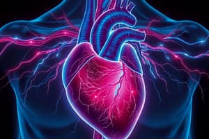
Cardiovascular System
Pulmonary and Systemic Circuits
- The right side of the heart pumps blood to the pulmonary circuit to pick up oxygen and dispose of carbon dioxide.
- The right atrium receives blood returning from the systemic circuit, and the right ventricle pumps blood into the pulmonary circuit.
- The left side receives oxygenated blood from the lungs and pumps it to the systemic circuit.
- The left atrium receives blood from the pulmonary circuit, and the left ventricle pumps blood into the systemic circuit.
Heart Location
- The heart is located obliquely within the mediastinum of the throax.
- It is mostly enclosed within this cavity and extends from the 2nd rib to the 5th intercostal space.
Heart Covering
- The heart is enclosed in a double-walled sac called the pericardium.
- The tough, dense connective tissue layer protects, anchors, and prevents overfilling.
- The serous pericardium is deep to this fibrous layer.
Serous Pericardium
- The serous pericardium is a thin, two-layered membrane forming a closed sac around the heart.
- Its parietal layer lines the fibrous pericardium.
- It also provides lubrication and protection.
- The visceral layer (epicardium) adheres to the surface of the heart.
- The pericardial cavity between layers contains serous fluid for lubrication.
Heart Wall Structure & Function
- Epicardium is the outermost layer that are made up of connective and epithelial tissues. It provides a smooth lubricated surface.
- Myocardium is the middle, contractile layer composed of cardiac muscles and fibrous skeleton.
- Endocardium is the innermost layer that is composed of the smooth endothelium in the inner myocardial surface.
Heart Chambers
- The heart has four chambers: right atrium and ventricle, left atrium and ventricle.
Atria Anatomy
- Atria receive returning blood and have smooth walls.
- Pectinate muscles form ridges in the anterior walls of right atrium.
- Fossa ovalis is found only in the left atria, and only in the auricle.
Blood Vessels & Atria
- The right atrium gets blood through the superior and the inferior vena cava.
- The coronary sinus drains the blood from the myocardium.
- Four veins carry oxygenized blood into the left atria.
Ventricle Anatomy
- Ventricle walls are more massive.
- Trabeculae carneae and papillary muscles mark the internal walls.
- The right ventricle is the anterior surface and pumps blood into the pulmonary trunk.
- The left ventricle dominates the posteroinferior surface and pumps blood into the aorta.
Ventricle Size & Blood flow
- The right ventricle is thinner-walled to pump deoxygenated blood under less pressure into the lungs.
- The left ventricle pumps oxygenated blood to the rest of the body at a greater pressure making it thicker.
Atrioventricular Valves
- The AV valves are located at each atrial-ventricular junction to prevent backflow into the atria in contractions..
- The right AV valve is tricuspid valve with three flexible cusps. The left is only bicuspid.
- The cusps are attached to chordae tendineae that are made of white collagin fibers.
- When the heart is relaxed, the AV valve flaps hang limply into the ventricle chambers with blood flowing through.
- When the ventricles compress the blood, the intraventricular pressure rises forcing blood to close them.
Papillary Muscles
- These are tethers that anchors the flaps to the ventricle walls.
- They tense to prevent flaps blowing upward in contractions.
Semilunar Valves
- The pulmonary valve is located at the junction of the right ventricle and pulmonary trunk.
- The aortic valve is at the junction of the left ventricle and aorta.
- The valves have 3 pocket-like cups that open and close upon pressure differences, which then either flatten against arteries as blood rushes thru or are pushed closed.
Coronary Circulation
Function
- This circulation is the functional blood supply for the heart.
- The heart has its own system for nourishment, where despite being filled by blood it runs on its own vessels to nourish it.
Arteries
- The LCA runs towards the heart dividing into the anterior interventricular region as well as the circumflex region.
- RCA rises to the lateral sides and supplies blood to two more branches.
- The Posterior region serves both ventricles
Veins
- Cardiac veins collect the blood and merge into the coronary sinus.
- Great and small cardiac veins drain into it.
- Veins collect oxygen poor blood.
Cardiac Muscle Anatomy
Structure
- Muscle is defined by striations and sliding filament mechanism
- cells are branched and interconnected with only one or two nuclei. It is abundant in the mitochondria
- Intracellular includes connective-tissue filled matrix for the fibers and its skeleton
- intercalated disks connect the muscle end to end with desmosomes and gap junctions Myofilbrils are composed or sarcomeres , made with A and I bands
Function
- matrix helps the cardiac cells exert force
- desmosomes helps keep adjacent cells together in contraction
- gap junctions allow ions to pass from cell to cell, helping them behave as one unit.
Skeletal vs Cardiac Muscle
- Skeletal : Individual simulated
- Cardiac: Gap junction
Tetanic contractions
- Skeletal: possible short refractionary period
- Cardiac: not possible long duration
Aerobic
- Skeletal: can create ATP aerobically.
- Cardiac: relies on aerobic
Ca+ sources
- Skeletal: is released and contracted.
- Cardiac: comes from the SR and extracellular fluid/regeneration
Slow Ca2+ Channels - Function & Importance
- Specialized channels in plasma membrane of cardiac cells.
- Their opening is much slower, that they trigger the release of ions need for muscular contraction.
- Calcium ion prolongs in the depolarization phase.
- It is used in contractions and calcium release of continuous activities
Intrinsic Conduction Systen
- The system consists of non contractible cardiac cells used to initiate the wave.
- The Sino-atrial (SA) node is located in the right atria.
- The Atrioventricular (AV) node is located in the inferior portion.
- Runs along the septum, dividing at the apex.
Conduction System Function
- Specialized cells generate and transmit impulses to the cardiac cells, creating a coordinated functions.
- The SA node initiates by generating action potentials around 75 beats.
- AV nodes delay impulses so the atria will fully contract before the next beat (roughly 0.1 sec)
- Dysfunctions with the heart result in irregular ventricular constrictors, that may require a pace maker.
- The purkinje fibers will deliver the impulse to help ensure coordination.
Different Types of Arrhythmias
- Fibrillation: disorganized contractions
- Extrasystole: Hyperexcitable parts cause premature constrictions.
- Heart Block: damaged nodes or branches can lead to blocked impulses.
Extrinsic Innervation
- The autonomy of the heart is modified
- The autonomic nervous system modified through each beat.
- The nervous system increases more frequently and forcefully.
- Activation slows the heart.
Pacemaker Cells
- Pacemaker potential is unstable, depolarized and has less sodium.
- Repolarizes after peak opens and closes.
Action Potentials vs Muscle/Never
- pacemaker cells have an unstable resting potential.
- calcium vs sodium
Heart Rhythm
- Diastole: is a resting peroid
- Systole: is the contraction period
Vavlves
Atrial Systole
- pressure slightly goes up as atria contracts
- AV valves remain open
- no change with valves
Ejection
- pressure peaks high at the the ventricles
- SL valves remain open.
- volume decreases
Ventrilicular finshing
a. Atrial pressure rises.
Factors of blood and the heart
- definition and rate
- reserve to push out over and over/reserve.
- Factors help each other out
Stroke Volume Defition
Factors and regulation
- Volume is key factor to regulate contractions
- SV represents the difference between the end and diastolic cycle.
- pressure of blood in ventricles
- Three factor are preload, contractions, afterload
Preload
- The degree to which muscle cells are stretched right before they contract
- Frank Starling Law helps increase the rate of increasing blood as well
Afterload
- Must always always beat to protect blood.
- Afterload and hypertension =less pressure of ventrices as well.
- More blood remains Factors to decrease
- Cold reduces hr
heart rate
tacycardia
- more than 100 bpm 1 May result from elevated body temp, stress, certain drugs, or heart disease
bradycardia.
1.May results with low temp, activates PNS
Heart issues
a. Coronary problems = poor delivery of oxygen b. BP = constant high c. Heart Attacks d. D = unknown reasons
Studying That Suits You
Use AI to generate personalized quizzes and flashcards to suit your learning preferences.