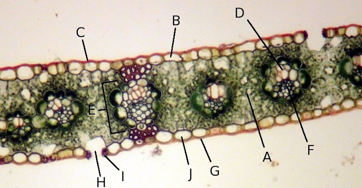
What are the labeled parts of the plant tissue in the image?

Understand the Problem
The question is likely related to identifying or explaining different parts of the plant tissue shown in the image. It appears to be a histological slide of plant anatomy, possibly asking for the functions or characteristics of labeled structures.
Answer
A - Xylem, B - Phloem, C - Epidermis, D - Palisade Mesophyll, E - Vascular Bundle, F - Spongy Mesophyll, G - Cuticle, H - Stomata, I - Guard Cells, J - Lower Epidermis.
The labeled parts of the plant tissue image are typically as follows: A - Xylem, B - Phloem, C - Epidermis, D - Palisade Mesophyll, E - Vascular Bundle, F - Spongy Mesophyll, G - Cuticle, H - Stomata, I - Guard Cells, J - Lower Epidermis.
Answer for screen readers
The labeled parts of the plant tissue image are typically as follows: A - Xylem, B - Phloem, C - Epidermis, D - Palisade Mesophyll, E - Vascular Bundle, F - Spongy Mesophyll, G - Cuticle, H - Stomata, I - Guard Cells, J - Lower Epidermis.
More Information
These labels represent common structures in a leaf cross-section, crucial for respiration, photosynthesis, and nutrient transport.
Tips
A common mistake is confusing xylem and phloem. Remember, xylem typically stains red and phloem stains green on color-prepped slides.
Sources
- 3.1.3: Plant Tissues - Biology LibreTexts - bio.libretexts.org
AI-generated content may contain errors. Please verify critical information