Podcast
Questions and Answers
Which of the following accessory organs is NOT directly part of the alimentary canal?
Which of the following accessory organs is NOT directly part of the alimentary canal?
- Liver
- Salivary glands
- Pancreas
- Esophagus (correct)
What is the primary function of the ileocecal sphincter?
What is the primary function of the ileocecal sphincter?
- Controlling the passage of food from the small intestine into the large intestine (correct)
- Facilitating the absorption of water in the large intestine
- Preventing the backflow of stomach contents into the esophagus
- Regulating the flow of chyme from the stomach into the duodenum
Which enzyme is primarily responsible for the initial breakdown of carbohydrates in the mouth?
Which enzyme is primarily responsible for the initial breakdown of carbohydrates in the mouth?
- Amylase (correct)
- Trypsin
- Pepsin
- Lipase
What is the role of hydrochloric acid (HCl) secreted in the stomach?
What is the role of hydrochloric acid (HCl) secreted in the stomach?
Which region of the small intestine is primarily responsible for the absorption of fats?
Which region of the small intestine is primarily responsible for the absorption of fats?
What is the main function of the microbiota bacteria found in the colon?
What is the main function of the microbiota bacteria found in the colon?
Which of the following enzyme-macronutrient pairings is INCORRECT?
Which of the following enzyme-macronutrient pairings is INCORRECT?
What hormone, released due to the presence of fat in the duodenum, signals the gallbladder to release bile?
What hormone, released due to the presence of fat in the duodenum, signals the gallbladder to release bile?
Which function of the GI tract involves the movement of nutrients and water from the lumen into the body?
Which function of the GI tract involves the movement of nutrients and water from the lumen into the body?
Which tissue layer of the GI tract contains the enteric neurons known as the 'gut brain'?
Which tissue layer of the GI tract contains the enteric neurons known as the 'gut brain'?
Which type of epithelium is found lining the esophagus?
Which type of epithelium is found lining the esophagus?
What is the primary function of foveolar cells in the stomach?
What is the primary function of foveolar cells in the stomach?
What structural feature of the small intestine significantly increases its surface area for absorption?
What structural feature of the small intestine significantly increases its surface area for absorption?
Which type of GI disorder is achalasia?
Which type of GI disorder is achalasia?
What is a common cause of peptic ulcers, a type of secretion disorder?
What is a common cause of peptic ulcers, a type of secretion disorder?
Which symptom is primarily associated with altered ingestion?
Which symptom is primarily associated with altered ingestion?
How do opioids reduce pain in the GI tract?
How do opioids reduce pain in the GI tract?
If a patient presents with black or tarry stool, where is the most likely location of bleeding in the GI tract?
If a patient presents with black or tarry stool, where is the most likely location of bleeding in the GI tract?
What is the PRIMARY function of the Upper Esophageal Sphincter (UES)?
What is the PRIMARY function of the Upper Esophageal Sphincter (UES)?
Which pathophysiological process is characteristic of achalasia?
Which pathophysiological process is characteristic of achalasia?
What is the primary mechanism by which proton pump inhibitors (PPIs) help in the treatment of GERD?
What is the primary mechanism by which proton pump inhibitors (PPIs) help in the treatment of GERD?
Which cell type is primarily responsible for secreting hydrochloric acid (HCl) in the stomach?
Which cell type is primarily responsible for secreting hydrochloric acid (HCl) in the stomach?
What is the primary function of Intrinsic Factor produced by the parietal cells in the stomach?
What is the primary function of Intrinsic Factor produced by the parietal cells in the stomach?
Which of the following mechanisms is involved in the protective actions of prostaglandins in the stomach?
Which of the following mechanisms is involved in the protective actions of prostaglandins in the stomach?
Which virulence factor of Helicobacter pylori disrupts actin, leading to inflammation?
Which virulence factor of Helicobacter pylori disrupts actin, leading to inflammation?
Which of the following types of stomach cancer arises from mucus-producing cells?
Which of the following types of stomach cancer arises from mucus-producing cells?
What is the function of the hepatic artery in supplying blood to the liver?
What is the function of the hepatic artery in supplying blood to the liver?
Hepatocytes have microvilli on their basal lateral side. Which of the following is a function of the hepatocyte?
Hepatocytes have microvilli on their basal lateral side. Which of the following is a function of the hepatocyte?
Which liver cell type is responsible for removing particulates from circulation?
Which liver cell type is responsible for removing particulates from circulation?
What is the role of sinusoidal endothelial cells in the liver?
What is the role of sinusoidal endothelial cells in the liver?
The liver performs important functions, but which of the following is a LIVER function, regarding protein metabolism?
The liver performs important functions, but which of the following is a LIVER function, regarding protein metabolism?
What is the primary transmission route for Hepatitis A?
What is the primary transmission route for Hepatitis A?
What is the cause of Non-Alcoholic Steatohepatitis (NASH)?
What is the cause of Non-Alcoholic Steatohepatitis (NASH)?
What process triggers the activation of stellate cells in the pathogenesis of liver cirrhosis?
What process triggers the activation of stellate cells in the pathogenesis of liver cirrhosis?
What component is lacking in the bile of patients with Cholestasis?
What component is lacking in the bile of patients with Cholestasis?
What is the primary component of Brown Pigment Gallstones?
What is the primary component of Brown Pigment Gallstones?
Why is Cholangitis so severe?
Why is Cholangitis so severe?
Where do the common bile duct and major pancreatic duct lead into?
Where do the common bile duct and major pancreatic duct lead into?
What is the role of secretin in the exocrine function of the pancreas?
What is the role of secretin in the exocrine function of the pancreas?
Which enzyme requires bile salts to function properly?
Which enzyme requires bile salts to function properly?
How do gallstones cause acute pancreatitis?
How do gallstones cause acute pancreatitis?
What are the primary symptoms associated with Cholangitis?
What are the primary symptoms associated with Cholangitis?
In type 1 diabetes, what component is responsible for activating dendritic cells to release corticotropin-releasing factor?
In type 1 diabetes, what component is responsible for activating dendritic cells to release corticotropin-releasing factor?
What distinguishes the jejunum from the duodenum and ileum?
What distinguishes the jejunum from the duodenum and ileum?
In the small intestine, M cells are MOST directly involved in:
In the small intestine, M cells are MOST directly involved in:
What is the primary role of bile salts in the digestion of fats within the small intestine?
What is the primary role of bile salts in the digestion of fats within the small intestine?
Which combination of enzymes and breakdown products is involved in protein digestion in the small intestine?
Which combination of enzymes and breakdown products is involved in protein digestion in the small intestine?
What structural feature significantly amplifies the surface area available for absorption in the small intestine?
What structural feature significantly amplifies the surface area available for absorption in the small intestine?
How do chylomicrons assist in the absorption of fats in the small intestine?
How do chylomicrons assist in the absorption of fats in the small intestine?
A patient experiences pain relief upon eating but later reports the pain returns with increased intensity. Which disorder is MOST likely to be the cause?
A patient experiences pain relief upon eating but later reports the pain returns with increased intensity. Which disorder is MOST likely to be the cause?
What is the primary mechanism behind the burning pain associated with duodenal peptic ulcers?
What is the primary mechanism behind the burning pain associated with duodenal peptic ulcers?
In Celiac disease, what component of gluten is MOST directly involved in triggering the associated autoimmune response?
In Celiac disease, what component of gluten is MOST directly involved in triggering the associated autoimmune response?
What is the role of tissue transglutaminase (tTG) in the pathogenesis of celiac disease?
What is the role of tissue transglutaminase (tTG) in the pathogenesis of celiac disease?
The disengagement of tight junctions in enterocytes, leading to increased intestinal permeability in celiac disease, is MOST directly caused by:
The disengagement of tight junctions in enterocytes, leading to increased intestinal permeability in celiac disease, is MOST directly caused by:
In lactose intolerance, the symptoms experienced by affected individuals are the direct result of:
In lactose intolerance, the symptoms experienced by affected individuals are the direct result of:
What is the primary absorptive function of the large intestine?
What is the primary absorptive function of the large intestine?
How does the microbiome in the large intestine contribute to human health?
How does the microbiome in the large intestine contribute to human health?
What is the main distinction between acute and chronic diarrhea?
What is the main distinction between acute and chronic diarrhea?
Why does fasting relieve osmotic diarrhea but not secretory diarrhea?
Why does fasting relieve osmotic diarrhea but not secretory diarrhea?
How do the sodium-glucose symporters facilitate water absorption in the large intestine?
How do the sodium-glucose symporters facilitate water absorption in the large intestine?
What is a common mechanism contributing to motility diarrhea?
What is a common mechanism contributing to motility diarrhea?
What is the primary effect of opiates and opioid derivatives as antidiarrheal medications?
What is the primary effect of opiates and opioid derivatives as antidiarrheal medications?
What is a common qualitative symptom of constipation?
What is a common qualitative symptom of constipation?
What is a key characteristic of Irritable Bowel Syndrome (IBS)?
What is a key characteristic of Irritable Bowel Syndrome (IBS)?
Corticotropin-releasing factor (CRF) has a direct role in the mechanisms of what disorder:
Corticotropin-releasing factor (CRF) has a direct role in the mechanisms of what disorder:
Which of the following is a distinguishing feature of Crohn's disease compared to ulcerative colitis?
Which of the following is a distinguishing feature of Crohn's disease compared to ulcerative colitis?
In a patient with altered gut function, which symptom is more indicative of ulcerative colitis than Crohn's disease?
In a patient with altered gut function, which symptom is more indicative of ulcerative colitis than Crohn's disease?
What is the BEST description of obesity?
What is the BEST description of obesity?
An individual with a high muscle mass is misclassified as overweight when only using BMI. Which of the following measurement provides the MOST accurate assessment for obesity in this case?
An individual with a high muscle mass is misclassified as overweight when only using BMI. Which of the following measurement provides the MOST accurate assessment for obesity in this case?
Which trend has been observed regarding obesity rates over time?
Which trend has been observed regarding obesity rates over time?
The 'Thrifty Gene Hypothesis' suggests that some genes:
The 'Thrifty Gene Hypothesis' suggests that some genes:
Which activity contributes the LEAST to total energy expenditure?
Which activity contributes the LEAST to total energy expenditure?
Which brain pathway involves the dopamine circuits in the substantia nigra sending signals to the motor cortex and promotes the desire for more dopamine through movement?
Which brain pathway involves the dopamine circuits in the substantia nigra sending signals to the motor cortex and promotes the desire for more dopamine through movement?
What is the role of glucagon-like peptide-1 (GLP-1) on long-term control of food intake?
What is the role of glucagon-like peptide-1 (GLP-1) on long-term control of food intake?
What is the difference between dieting and Bariatric Surgery on hormones related to hunger?
What is the difference between dieting and Bariatric Surgery on hormones related to hunger?
When evaluating a patient for possible GERD, which symptom is MOST indicative but requires excluding cardiac issues first?
When evaluating a patient for possible GERD, which symptom is MOST indicative but requires excluding cardiac issues first?
What findings regarding tests on liver injury show the MOST indication of biliary obstruction?
What findings regarding tests on liver injury show the MOST indication of biliary obstruction?
A patient with chronic liver disease has decreased synthesis of serum albumin. What condition does this trigger?
A patient with chronic liver disease has decreased synthesis of serum albumin. What condition does this trigger?
A patient with Gilbert's syndrome may manifest what visible symptom, related to liver function?
A patient with Gilbert's syndrome may manifest what visible symptom, related to liver function?
What liver test pattern is MOST directly indicative of biliary tract damage?
What liver test pattern is MOST directly indicative of biliary tract damage?
The jejunum is MOSTLY responsible for what function?
The jejunum is MOSTLY responsible for what function?
Which layer of the GI tract contains blood and lymphatic vessels, as well as glands?
Which layer of the GI tract contains blood and lymphatic vessels, as well as glands?
What is the PRIMARY function of the mucus secreted by goblet cells in the colon?
What is the PRIMARY function of the mucus secreted by goblet cells in the colon?
What is the initial response when food enters the stomach in the Gastric Phase of acid secretion?
What is the initial response when food enters the stomach in the Gastric Phase of acid secretion?
Which of the following is the correct order of histological progression to cancer in prolonged GERD?
Which of the following is the correct order of histological progression to cancer in prolonged GERD?
What is a key pathological feature that OVERLAPS between peptic ulcers caused by NSAIDs and those caused by H. pylori infection?
What is a key pathological feature that OVERLAPS between peptic ulcers caused by NSAIDs and those caused by H. pylori infection?
What is the MOST LIKELY consequence of a loss of intrinsic factor production in the stomach?
What is the MOST LIKELY consequence of a loss of intrinsic factor production in the stomach?
Damage to the vagus nerve would MOST directly affect which phase of acid secretion in the stomach?
Damage to the vagus nerve would MOST directly affect which phase of acid secretion in the stomach?
What is a PRIMARY mechanism by which alcohol contributes to liver cirrhosis?
What is a PRIMARY mechanism by which alcohol contributes to liver cirrhosis?
What is the MOST DIRECT impact of cirrhosis on drug metabolism and availability?
What is the MOST DIRECT impact of cirrhosis on drug metabolism and availability?
What is the BEST course of action if a cirrhotic patient exhibits ascites?
What is the BEST course of action if a cirrhotic patient exhibits ascites?
Which set of conditions defines Charcot's Triad, MOST indicative of Cholangitis?
Which set of conditions defines Charcot's Triad, MOST indicative of Cholangitis?
What is the MOST SIGNIFICANT risk associated with gallstones blocking the ampulla of Vater?
What is the MOST SIGNIFICANT risk associated with gallstones blocking the ampulla of Vater?
What is the MAIN effect of secretin on the exocrine pancreas?
What is the MAIN effect of secretin on the exocrine pancreas?
What is the primary process causing auto digestion in acute pancreatitis?
What is the primary process causing auto digestion in acute pancreatitis?
In Celiac disease, which factor contributes to the loss of enterocyte integrity?
In Celiac disease, which factor contributes to the loss of enterocyte integrity?
Patients with diarrhea caused by increased luminal osmolarity experience relief with fasting because:
Patients with diarrhea caused by increased luminal osmolarity experience relief with fasting because:
How does Corticotropin-Releasing Factor (CRF) impact Irritable Bowel Syndrome (IBS)?
How does Corticotropin-Releasing Factor (CRF) impact Irritable Bowel Syndrome (IBS)?
Which of the following is NOT a long-term adjustment related to Bariatric Surgery?
Which of the following is NOT a long-term adjustment related to Bariatric Surgery?
Which test is most specific to the liver for detecting injury?
Which test is most specific to the liver for detecting injury?
Flashcards
GI Tract
GI Tract
Hollow tube from mouth to anus, includes accessory organs.
Mouth's Role in Digestion
Mouth's Role in Digestion
Chews and mashes food; saliva releases amylase and lipase
Esophagus Function
Esophagus Function
Transports food bolus to stomach
Stomach's Role in Digestion
Stomach's Role in Digestion
Signup and view all the flashcards
Duodenum Function
Duodenum Function
Signup and view all the flashcards
Jejunum and Ileum
Jejunum and Ileum
Signup and view all the flashcards
Colon Function
Colon Function
Signup and view all the flashcards
Rectum and Anus Function
Rectum and Anus Function
Signup and view all the flashcards
Amylase Role
Amylase Role
Signup and view all the flashcards
Pepsin, Trypsin, Chymotrypsin
Pepsin, Trypsin, Chymotrypsin
Signup and view all the flashcards
Lipase Role
Lipase Role
Signup and view all the flashcards
GI Tract Functions
GI Tract Functions
Signup and view all the flashcards
Cholecystokinin (CCK)
Cholecystokinin (CCK)
Signup and view all the flashcards
Epithelium
Epithelium
Signup and view all the flashcards
Lamina Propria
Lamina Propria
Signup and view all the flashcards
Muscularis Mucosa
Muscularis Mucosa
Signup and view all the flashcards
Submucosa Layer
Submucosa Layer
Signup and view all the flashcards
Muscularis Layer
Muscularis Layer
Signup and view all the flashcards
Myenteric Plexus
Myenteric Plexus
Signup and view all the flashcards
Serosa Layer
Serosa Layer
Signup and view all the flashcards
Tunica Mucosa
Tunica Mucosa
Signup and view all the flashcards
Esophagus Epithelium Function
Esophagus Epithelium Function
Signup and view all the flashcards
Stomach Epithelium Function
Stomach Epithelium Function
Signup and view all the flashcards
Small Intestine Epithelium
Small Intestine Epithelium
Signup and view all the flashcards
Colon Epithelium Function
Colon Epithelium Function
Signup and view all the flashcards
Motility Disorders
Motility Disorders
Signup and view all the flashcards
Secretion Disorders
Secretion Disorders
Signup and view all the flashcards
Digestion Disorders
Digestion Disorders
Signup and view all the flashcards
Absorption Disorders
Absorption Disorders
Signup and view all the flashcards
Dysphagia
Dysphagia
Signup and view all the flashcards
Odynophagia
Odynophagia
Signup and view all the flashcards
Anorexia
Anorexia
Signup and view all the flashcards
Ascending Pain Pathway
Ascending Pain Pathway
Signup and view all the flashcards
Descending Pain Pathway
Descending Pain Pathway
Signup and view all the flashcards
Opioids Action
Opioids Action
Signup and view all the flashcards
NSAIDs Action
NSAIDs Action
Signup and view all the flashcards
Visceral Pain
Visceral Pain
Signup and view all the flashcards
Nausea
Nausea
Signup and view all the flashcards
Vomiting
Vomiting
Signup and view all the flashcards
Chemoreceptor Trigger Zone
Chemoreceptor Trigger Zone
Signup and view all the flashcards
Muscarinic Receptors
Muscarinic Receptors
Signup and view all the flashcards
5-HT3 Receptor Antagonists
5-HT3 Receptor Antagonists
Signup and view all the flashcards
Pharyngeal Phase
Pharyngeal Phase
Signup and view all the flashcards
Esophageal Phase
Esophageal Phase
Signup and view all the flashcards
Esophagus Definition
Esophagus Definition
Signup and view all the flashcards
Upper Esophageal Sphincter (UES)
Upper Esophageal Sphincter (UES)
Signup and view all the flashcards
Lower Esophageal Sphincter (LES)
Lower Esophageal Sphincter (LES)
Signup and view all the flashcards
Peristalsis
Peristalsis
Signup and view all the flashcards
Achalasia Definition
Achalasia Definition
Signup and view all the flashcards
Study Notes
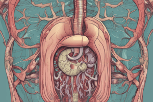
Overview of the GI System
- The GI tract is a complex, multi-organ system composed of the alimentary canal, a hollow tube from the mouth to the anus, along with accessory organs like salivary glands, liver, gallbladder, and pancreas.
- It is 7-9 meters long in an adult human.
- It includes the mouth, esophagus, stomach, small intestine (duodenum, jejunum, ileum), large intestine (colon, cecum), rectum, and anus.
Digestion Process
Mouth
- Food is mechanically chewed, mixed, and mashed with saliva.
- Salivary glands release amylase (breaks down carbohydrates) and lipase (breaks down fat).
Esophagus
- The food bolus passes through the upper esophageal sphincter, down the esophagus, past the lower esophageal sphincter, and into the stomach.
Stomach
- Mechanical contractions mix the food.
- Parietal cells secrete hydrochloric acid.
- Chief cells secrete pepsin (breaks down proteins) and lipase (breaks down fat).
- Food becomes a liquid solution called chyme.
Small Intestine
Duodenum
- Chyme passes through the pyloric sphincter into the duodenum.
- Digestion occurs with contractions and enzymes from epithelial cells.
- Bile from the gallbladder and liver mixes with chyme.
- The pancreas secretes amylase, trypsin, chymotrypsin, and lipase.
Jejunum and Ileum
- Bulk absorption and reabsorption occurs.
- Nutrients and most water are reabsorbed.
- The ileum absorbs more fats.
- The jejunum absorbs more carbohydrates and amino acids.
Large Intestine
Cecum
- The food bolus passes through the ileocecal sphincter and rests briefly in the cecum.
Colon
- More water is absorbed.
- Microbiota bacteria help break down fibers.
Rectum and Anus
- Food waste is stored in the rectum and passes through the anus.
Macronutrient Breakdown
- Carbohydrates are broken down by amylase in the mouth, duodenum (via pancreas), and jejunum.
- Proteins are broken down by pepsin, trypsin, and chymotrypsin in the stomach and duodenum (via pancreas).
- Fats are broken down by lipase in the mouth, stomach, duodenum (via gallbladder/liver/pancreas), and ileum.
Functions of the GI Tract
- Digestion: Breakdown of food.
- Secretion of hormones: Exocrine and endocrine secretion.
- Absorption: Nutrients and water from the lumen into the body.
- Motility: Mixing and movement of food.
- Defense: Barrier against harmful components; largest immune organ.
- Gut microbiome: Breaks down fibers, aids in digestion, and affects diseases.
- Exocrine secretion releases enzymes into the digestive tract for chemical digestion.
- Endocrine secretion helps coordinate digestion events, glucose regulation, and hunger. Example: CCK.
- CCK is released from the duodenum due to fat, enters the bloodstream, binds to receptors in the gallbladder to signal the release of bile and inhibits the opening of the pyloric sphincter.
GI Anatomy - Tissue Layers
Tunica Mucosa
- Epithelium: Cells lining the lumen.
- Lamina Propria: Connective tissue with immune cells.
- Muscularis Mucosa: Thin muscle layer providing structure and aiding in secretion.
Submucosa Layer
- Dense connective tissue connecting glands, blood, and lymphatic tissue.
Muscularis Layer
- Circular and longitudinal smooth muscle layers.
- Myenteric Plexus: Enteric neurons between smooth muscle layers (the "gut brain").
Serosa Layer
- Mesothelium that lines the organ.
Histology of GI Tract
Esophagus
- Stratified squamous epithelium.
- Deals with damage from swallowing; multiple layers of square, flat cells that slough off when damaged by food.
Stomach
- Simple columnar epithelium that forms deep pits.
- Specialized epithelial cells secrete acids and enzymes.
- Foveolar cells produce mucus to protect from acid.
Small Intestine
- Single columnar epithelium forming villi.
- Enterocytes with microvilli create a brush border.
- The brush border is important for reabsorption and provides a large surface area.
- Goblet cells produce mucus.
Colon
- Single columnar epithelium organized in a flat surface.
- Predominantly goblet cells that produce mucus.
- Absorptive enterocytes reabsorb water.
Overview of GI Disorders
- Categorized into motility, secretion, digestion, or absorption disorders.
Motility Disorders
- Affect all major regions of the GI tract.
- Examples: Achalasia or Hirschsprung's disease prevent proper smooth muscle contraction.
Secretion Disorders
- Inappropriate production of acid or mucus in the stomach, digestive enzymes, or bicarbonate from the pancreas, or too much/little bile from the liver.
- Example: Peptic ulcers result from secreting too much hydrochloric acid.
Digestion Disorders
- Incomplete breakdown of nutrients.
- Example: Lactose intolerance.
Absorption Disorders
- Decreased ability to absorb nutrients.
- Example: Crohn's disease.
GI Disease Symptoms
- Pain
- Altered Ingestion: Nausea or vomiting, dysphagia (difficulty swallowing), odynophagia (painful swallowing), anorexia (lack of appetite).
- Altered Bowel Movements: Diarrhea or constipation.
- GI Tract Bleeding
- These are symptoms of the problem, not the problem themselves.
Pain Pathways
Ascending Pathway
- Damage sends a message to the dorsal root ganglia (DRG) outside the spinal cord.
- Message transmits to neurons in the spinal cord.
- Neurons travel up the spinal cord to the thalamus.
- The thalamus connects with the somatosensory cortex, signaling pain.
Descending Pathway
- Inhibits the signal at the level of the spinal cord, which reduces pain signals.
Opioids
- Inhibit the ascending pathway at the dorsal root ganglion and spinal cord.
- Activate the descending pathway to increase inhibition.
Non-Steroidal Anti-Inflammatory Drugs (NSAIDs)
- Act at the site of damage to reduce inflammation, which reduces signals sent to the brain.
Visceral Pain
- Poorly localized pain often referred to different structures.
- Referred pain theory: First-order neurons from different organs synapse on the same second-order neuron in the spinal cord, so the brain cannot differentiate the origin of the pain.
Nausea and Vomiting
- Nausea: The feeling of sickness in the stomach with an inclination to vomit.
- Vomiting: Forcible ejection of undigested GI content from the mouth and a defense mechanism.
- Relaxation of the lower esophageal sphincter.
- Increased intra-abdominal pressure.
- Contraction of diaphragm and abdominal muscles.
- Closing of the epiglottis.
Vomiting Reflex Mechanism
Brainstem
- Vomiting center (muscarinic receptors trigger the vomiting reflex).
- Chemoreceptor trigger zone (located outside the blood-brain barrier).
Chemoreceptor Trigger Zone
- Detects toxins from chemotherapy, radiation, or ingested toxins.
- Triggers vomiting reflex.
Higher Brain Centers
- Sensory, pain, smell, sight, memory, fear, or anticipation can trigger the vomiting center.
Gut Signals
- Food that is not digested properly sends a signal to the vagus nerve.
- The vagus nerve signals the vomiting center.
Medications to Treat Nausea and Vomiting
- 5-HT3 Receptor Antagonists (Ondansetron/Zofran): Commonly prescribed, relatively safe anti-vomiting medication.
- Prochlorperazine: Anti-vomiting and antipsychotic drug (D2 receptor antagonist).
- Scopolamine: Muscarinic receptor antagonist used for motion sickness (prescribed as a patch).
Dysphagia and Odynophagia
- Dysphagia: Difficulty swallowing.
- Odynophagia: Painful swallowing.
Phases of Swallowing
- Voluntary Phase: The bolus of food is pushed by the tongue past the hard palate toward the back of the throat.
- Pharyngeal Phase: Involuntary movement of the bolus into the oropharynx and soft palate of the nasopharynx, eliciting closure of the epiglottis.
- Esophageal Phase: Involuntary contraction of the esophagus muscles pushes the bolus down to the stomach.
Problems with Swallowing
- Neurological disorders may affect the voluntary phase.
- Stroke may affect the pharyngeal phase.
- GERD, achalasia, or esophageal cancer may affect the esophageal phase.
Anorexia
- Loss of desire to eat.
Primary Anorexia (Anorexia Nervosa)
- A neural disorder where the brain signals to stop eating.
Secondary Anorexia
- A consequence of a disease such as GERD, peptic ulcer, Crohn's disease, ulcerative colitis, or cholecystitis.
Potential Treatment
- CGRP receptor antagonist may be useful in treating anorexia by targeting hunger signals in the parabrachial nucleus.
Gastrointestinal Tract Bleeding
- The location and color of blood help in diagnosis.
- Bright Red Blood Coating the Stool: Bleeding at the rectum or anus; possible causes include hemorrhoids or polyps (especially in children).
- Dark Blood Mixed in the Stool: Colon problem.
- Black or Tarry Stool: Bleeding in the esophagus, stomach, or duodenum.
- Bright Red Blood in Vomit: Bleeding in the esophagus or stomach.
- Coffee Grounds Appearance in Vomit: Bleeding in the stomach or duodenum.
- Signs of Fatigue or Anemia: May indicate GI tract bleeding even without visible blood.
Study Guide Highlights
- Anatomical specializations that enable function along the length of the GI tract.
- Main functions of the GI tract.
- Main symptoms of GI disorders.
- Mechanisms of action of drugs for treating the main symptoms of GI disorders.
- Focus on the vomiting pathway and the pain pathway.
Anatomy and Function of the Esophagus
Basic Anatomy
- The esophagus connects the oral cavity to the stomach; it is located behind the trachea and in front of the spine.
Sphincters
- Upper Esophageal Sphincter (UES): Protects the airways.
- Lower Esophageal Sphincter (LES): Keeps food in the stomach and prevents acid reflux.
Function
- Transports swallowed food to the stomach.
- The UES allows food entry into the esophagus and protects the airway from swallowed material and gastric reflux. The epiglottis closes as the UES opens.
- The esophageal tube transports the food bolus from the pharynx to the stomach and clears refluxed material from the stomach.
- The mucosa cells and stratified squamous structure lining protect the esophagus from jagged food edges and stomach acid.
- The LES allows food entry into the stomach and protects the esophagus from gastric reflux.
Propulsive Actions (Peristalsis)
- The esophagus moves food via peristalsis (wave-like motion of contraction and relaxation); higher pressure indicates contraction.
Overview of Esophageal Disorders
- Achalasia: Over contraction of the lower esophageal sphincter.
- GERD: Reduced contraction of the lower esophageal sphincter.
Achalasia
Definition
- A motility disorder caused by incomplete LES relaxation.
Mechanism
- Esophageal sphincter will not relax.
- Dilation of the esophagus and food buildup at the LES.
Pathophysiology
- Inflammation of the Myenteric Plexus: Inflammation of neurons between smooth muscle layers can lead to loss of relaxation control over the sphincter muscle.
- Genetic Predisposition: May increase the risk of developing Achalasia if exposed to a virus that causes inflammation of the myenteric plexus.
- Reduction in Nitric Oxide Synthase: Autoimmune cells target dilating neurons, reducing nitric oxide, which is needed to relax smooth muscle.
Treatments
- Pharmacological treatments are not very effective.
- Best way to manage LES is surgery.
- Calcium Channel Blockers, Nitric Oxide Donors, and PD Inhibitors: Relax smooth muscle cells but are not specific to the sphincter.
- Surgery to Widen LES: A complication of surgery is the LES may not contract enough, leading to GERD.
Cause & Mechanism
- Inflammation of Myenteric Plexus: A virus or toxin can cause inflammation of the neurons located between the smooth muscle layers, leading to a loss of relaxation control over the sphincter muscle.
- Genetic Predisposition: A genetic component may increase the risk of developing Achalasia if exposed to a virus that causes inflammation of the myenteric plexus.
- Reduction in Nitric Oxide: Cytotoxic autoimmune cells target dilating neurons, reducing nitric oxide and preventing smooth muscle relaxation.
GERD
Definition
- Gastroesophageal reflux is when stomach contents come back up into the esophagus or beyond.
- GERD involves acid reflux, heartburn and chest pain at least weekly for three consecutive weeks.
Mechanism
- Lower esophageal sphincter does not constrict enough.
Symptoms
- Typical: Heartburn, chest pain, acid regurgitation, sour taste in the mouth, altered ingestion.
- Atypical: Epigastric pain, nausea, bloating, belching, bleeding. Reduced LES tone and increased gastric pressure lead to reflux into the esophagus.
Symptoms Chart
| Typical | Atypical | |
|---|---|---|
| Pain | Heartburn, chest pain | Epigastric pain |
| Ingestion | Altered ingestion | Nausea, bloating, belching |
| Other | Acid regurgitation, sour taste in mouth | Bleeding |
Treatments
- Lifestyle Modifications: weight loss, stop smoking, small and regular meals, avoiding meals prior to sleep, avoiding certain medications, carbonated drinks, caffeine, alcohol, citrus or spicy foods.
- Pharmacological Treatment:
- Proton Pump Inhibitors (PPIs): Inhibit the hydrogen potassium pump in parietal cells, reducing hydrochloric acid secretion.
- Antacids: Directly balance hydrochloric acid.
- H2 Receptor Antagonists: Block histamine receptors on parietal cells, reducing hydrochloric acid secretion.
- Things that might cause increased acid and pepsin: Certain medications, NSAIDs, highly acidic food, Alcohol, Large fatty meals that are difficult to digest (can have delayed gastric emptying), obesity, carbonated drinks, pregnancy (can lead to increased gastric pressure and can directly reduce the LES tone).
Disease Progression of GERD
- Delayed gastric emptying, increased frequency of LES relaxations, increased acidity, loss of secondary peristalsis, and decreased LES tone can lead to esophageal lesions.
- Recurrent injury and scarring can cause stricture pain, obstruction, and perforation.
- Repeated injury may lead to Barrett's esophagus and cancer. | Stage | Discription | | ------------- |:-------------:| |Normal Squamous Cell Layer | Normal esophageal lining | |Injury | Damage to te squamous cell layer| |Columnar Layer (Barrett's Esophagus) | Change to columnar epithelium| |Barrett's Esophagus with Dysplasia | Rapidly changing cells that shouldn't be there| |Adenocarcinoma | Cancer due to constant cell changes|
Complications of Prolonged GERD
Erosive Esophagitis
- Inflammation in the epithelium.
Peptic Strictures
- Narrowing of the esophagus from scarring tissues.
Barrett's Esophagus
- Metaplasia where stratified squamous epithelium turns into columnar epithelium, increasing the risk of esophageal adenocarcinoma; less than 20% survive beyond five years.
Histological Progression to Cancer
- Normal squamous cells transition to columnar cells (Barrett's esophagus), leading to dysplasia and adenocarcinoma.
Treatment
- Surgical removal of the esophagus with vagal sparing.
Alarm Systems
- Constant pain: Indicative of erosive damage.
- Dysphagia: Difficulty swallowing, indicative of strictures.
- GI bleeding or anemia.
- Weight loss: Indicative of adenocarcinoma.
Study Guide Highlights
- Function of the esophagus and how achalasia affects it.
- Common conditions for GERD and how progression changes the esophagus.
- Treatment symptoms of GERD and how they target the mechanisms.
Stomach Anatomy
- Esophagus connects to the lower esophageal sphincter.
- The lower esophageal sphincter goes into the cardia.
- The fundus is at the top of the stomach.
- The body is the main central region.
- The antrum leads to the pyloric sphincter.
- The pyloric sphincter controls the passage of chyme into the duodenum.
- Rugae: Folds of mucosa that allow the stomach to distend.
- Gastric pits: Indentations in the stomach wall that are the entrance to gastric glands.
Stomach Function
- Digestion: Mechanical and chemical breakdown of food. Contractions mix food with hydrochloric acid, pepsin, and lipases.
- Storage: Holds food while chyme moves through the duodenum.
- Intrinsic Factor Production: Parietal cells produce intrinsic factor, a glycoprotein necessary for vitamin B12 absorption; lack of intrinsic factor can lead to pernicious anemia due to impaired vitamin B12 absorption in the ileum.
Histology
- Cell Turnover: High turnover rate due to the acidic environment.
- Stem Cells: Located in the pits; continuously produce new calls to replace damage in the pylorus, body, & fundus by stomach acid.
- Secreting Cells: Parietal cells, ECL cells, and chief cells.
- Pylorus Protection: Contains more gland mucus cells, neck mucus cells, and surface mucus cells to protect against the acidic chyme.
- G Cells and D Cells: Also present in the pylorus. |Cell Type| Secretions| Function| |:----|:----|:----| |Foveolar Cells| Mucus, bicarbonate, surfactant| Protects the stomach lining.| |Parietal Cells| Hydrogen ions (H+) and chloride ions (Cl-)| Forms hydrochloric acid (HCl) for digestion.| |Chief Cells| Pepsinogen, gastric lipase|Pepsinogen breaks down peptides; gastric lipase breaks down lipids.| |ECL Cells| Histamine|Stimulates parietal cells to secrete HCl.| |G Cells|Gastrin|Stimulates acid secretion and gastric motility.| |D Cells|Somatostatin|Inhibits acid secretion.|
Regulation of Parietal Cells
Activation
- Gastrin.
- Parasympathetic nervous system.
- Histamine.
Inhibition
- Somatostatin (secreted by D cells).
Luminal vs. Basal Lateral Side
- Lumen: The inside of the GI tract tube, considered outside the body.
- Basal Lateral Side: The backside of the tube, facing the body's interior, absorption occurs from the lumen to the basal lateral side, entering the body.
Regulation of Acid Secretion
Cephalic Phase
- Thinking about, seeing, or smelling food triggers the vagus nerve to prepare the stomach for digestion.
Gastric Phase
Food Enters the Stomach
- Food enters the stomach, causing distension activating the vagal reflex.
- Acid buffers the food, increasing pH, preventing somatostatin secretion.
- The vagus nerve releases acetylcholine, directly activating parietal cells to secrete acid
- Acetylcholine stimulates ECL cells to release histamine, G cells release gastrin, both leading to increased HCL secretion.
- Amino acids activate G cells (gastrin secretion, HCL secretion).
- High HCL levels activate D cells to secrete somatostatin (inhibit acid secretion)
Protective Mechanisms
- Regulation of Acid and Enzyme Production: Released in response to food, especially amino acids.
- Feedback Mechanisms: Prevent overproduction of acid.
Mucosal Barrier
- Apical membrane resistance and gastric surfactant reduces surface tension to allow distension without damage.
- Mucus protects against hydrogen ions.
- Bicarbonate buffers hydrogen ions; both are secreted by mucosal cells.
Epithelial Regeneration
- Constant turnover of cells to protect against acid damage.
Vascularization
- Close proximity of blood vessels to the basal lateral membrane for fast absorption.
Gastric Disorders
Peptic Ulcer Disease (PUD)
- Definition : Break in the mucosal surface that reaches the submucosal layer.
- Cause: Constant inflammation (gastritis is due to excess acid or stress, impairs regeneration.
- Prevalence: Affects 10% of the population.
Symptoms:
- Burning pain in the upper middle abdomen; Worsens at night and in the early morning, and is relieved by eating.
- Severe chronic pain, nausea, altered appetite, and bloating
- Lack of appetite, significant weight loss, bleeding, black tarry stools, and vomiting blood is alarming symptoms.
Causes
- Helicobacter pylori infection.
- Excessive NSAID consumption.
- Stress.
- Genetic predisposition.
Helicobacter pylori
- Rod-like, gram-negative bacteria with flagella and BabA.
- Secretes urease to create a basic environment to protect from stomach acid.
- Virulence factors include CagA and VacA Mechanism of H. pylori:
Feeds on surfactant Generates ammonia using urease (basic environment) Flagella propels to epithelial layer. BabA: adhers to epithelial cells and injects CagA, disrupting actin. Secretes VacA, leading to apoptosis.
- Acid increases inflammation, damaging mucosa and submucosal layers.
NSAIDS
- COX inhibitors prevent the conversion of arachidonic acid to prostaglandins.
- Prostaglandins have cytoprotective effects, inhibit gastric acid production, promote inflammation and pain; prevent bleeding. Mechanism:
Disrupt protective mechanisms, cytotoxic to epithelial cells, reduce mucus production; reduce epithelial repair. Increase bleeding and acid production
Complications
- Hemorrhage: Blood vessel damage, coffee ground vomitus or tarry stools
- Perforation: Erosion through the entire wall; Leads to septic shock
- Obstruction: Swelling prevents food from leaving the stomach
- Increased risk of gastritis and gastric cancer
Gastritis
- Inflammation of the gastric lining, leading to a breakdown of the mucosal barrier.
Causes
- Toxic substances such as, alcohol, acids and bases.
- Reduced mucus synthesis, common in elderly.
- Infections, like H. Pylori
Symptoms
- Pain: Epigastric discomfort
- Altered ingestion : Anorexia, Burping
- Nausea, vomiting, bleeding, presence of melena or hematemesis
Complications
- Increased risk of peptic ulcer disease
- Severe bleeding or anemia
- Increased risk of stomach cancer
Stomach Cancer
Zollinger-Ellison Syndrome
- Gastrin-producing tumor (gastrinoma) in the antrum, duodenum, or pancreas
- Sporadic or caused by MEN-1 gene mutations
- Overproduction of hydrochloric acid, no response to somatostatin
- Treated with proton pump inhibitors and antacids; surgery should be performed for sporadic gastrinomas Symptoms:
Epigastric pain relieved by eating; diarrhea due to acid hypersecretion; nausea, vomiting, loss of appetite, and weight loss. Bleeding from the stomach at the site of the tumor
Types of Stomach Cancers
- Adenocarcinoma (mucus-producing cells).
- Lymphoma (overproduction of lymphocytes, usually B cells).
Risk Factors:
Increased risk after age 55.
Locations of Stomach Cancer
- Cardia :10%
- Fundus and body: 10%
- Greater curvature: 3-5%
- Lesser curvature: 25%
- Pyloric area: 50%
Symptoms
- Often asymptomatic until advanced
- Severe, persistent heartburn/stomach pains
- Persistent nausea, lack of appetite; Weight loss
- Chronic vomiting of blood, blood in stool Mechanism:
H. Pylori causes Chronic active Gastritis Immune response can be due to diet, genetics, and excess alcohol. This can cause atrophy (decrease loss of gastric glands).
Treatment
- Surgical removal, antibiotics for H. Pylori, chemotherapy, and radiotherapy
Study Guide Highlights
- Anatomical differences in epithelium between the body and the antrum.
- Protective barriers and how they are disrupted in peptic ulcer disease.
- Treatment of Zollinger-Ellison syndrome.
- Pathophysiology of gastric cancer.
Liver Anatomy and Function
- Two Lobes: The liver consists of a large right lobe and a smaller left lobe, covering part of the stomach.
- Connection to Biliary System: Connected to the gallbladder and duodenum via the bile duct and gallbladder bile duct.
Blood Supply
- Portal Vein: Carries 70% of venous blood from the esophagus to the rectum, transporting nutrients and toxins from the digestive system for liver processing.
- Hepatic Artery: Carries 30% of arterial (oxygenated) blood to supply the liver tissues.
Structural Organization of the Liver
-
Hepatic Lobules: Hexagonal-shaped structures containing a central vein.
-
Hepatic Triad: Located at the corners of each hexagon, consisting of an artery, a vein, and a bile canaliculi. |Component|Description|Function| |:----|:----|:----| |Portal Vein|Carries 70% of blood from the hepatic portal vein system into the central vein.|Transports nutrients and toxins from the digestive system to the liver for processing.| |Hepatic Artery|Carries oxygenated blood from the heart, mixing with blood from the portal vein in the central vein.|Supplies oxygen to the liver tissues.| |Bile Canaliculi| Transports bile produced by hepatocytes, flowing into the bile duct towards the duodenum and gallbladder.|Collects and transports bile, which aids in the digestion and absorption of fats.| |Hepatocytes|Square cells that produce bile.| Production of bile.| |Sinusoid|A large compartment where venous and arterial blood mixes, facilitating nutrient, peptide, and gas exchange, similar to a capillary.| Facilitates the exchange of nutrients, oxygen, and large proteins between the blood and liver cells.| |Endothelial Cell|Covers the sinusoid.| Forms the barrier of the sinusoids with large fenestrations, allowing large proteins to pass through.|
-
Sinusoids have larger openings for large protiens to enter the central vein and circulatory systems.
Liver Cells
| Cell Type | Location | Function |
|---|---|---|
| Hepatocytes | Main liver cells with microvilli on the basal lateral side (facing blood). | Metabolism of lipoproteins, bile, and proteins; drug detoxification. |
| Liver Sinusoidal Endothelial Cell | Line the barrier of the sinusoids. | Forms the barrier of the sinusoids with large fenestrations, allowing large proteins like albumin to move in and out. |
| Kupffer Cells | Fixed in the sinusoidal vascular space. | Liver macrophages that remove particulates from circulation. |
| Pit Cells | Liver-associated natural killer cells. | Part of the innate liver immunity and host defense. |
| Stellate Cells | Located in the space of Disse, between hepatocytes' microvilli and sinusoid. | Central role in vitamin A storage and pathogenesis of cirrhosis; produce fibrotic factors. |
- Kupffer Cells Detoxify the liver.
- pit cells kill natural killer cells
Major Functions of the Liver
- Processing Nutrients: Processes nutrients absorbed from the digestive system and releases them into circulation.
- Clearing Toxic Chemicals: Removes harmful and toxic chemicals from the body.
- Synthesis of Plasma Proteins: Synthesizes albumin, which is crucial for blood homeostasis, concentration, hormone transport, and blood viscosity.
- Production of Bile Salts: Aids in the digestion and absorption of fats.
Carbohydrate Metabolism
- Glycogenesis: Storage of glucose as glycogen.
- Glycogenolysis: Breakdown of glycogen into glucose.
- Gluconeogenesis: Formation of glucose.
Protein Metabolism
- Amino acids from the gut are deaminated to form ATP or glucose via gluconeogenesis.
Lipid Metabolism
- Bile acids aid in fat digestion.
- Fatty acids are absorbed by hepatocytes and metabolized into ATP or converted to glucose via gluconeogenesis, and can be stored as fat.
Protection and Clearance
- Kupffer cells: Phagocytize toxins and bacteria from the GI tract.
Detoxification
- Phase one involves oxidation of toxins to make them soluble.
- Phase two involves conjugation to glutathione for excretion through bile and urine.
- The liver also breaks down alcohol, drugs, and environmental chemicals into inactive metabolites.
Metabolic End Products
- Ammonia, urea, and bacterial endotoxins are filtered out to the kidney.
Synthesis
- Albumin: Maintains oncotic pressure and acts as a carrier for hormones
- Coagulation Factors: Fibrinogen and prothrombin are necessary for blood clots affect hematocrit
Iron Transport and Binding
- Apoferritin for iron storage and transferrin for iron transport
Liver Diseases
- Hepatitis (acute, chronic, cirrhosis).
- Cholestasis (gallstones).
- Liver cancer.
Hepatitis
- Acute Hepatitis: Inflammation of the liver, usually caused by hepatitis viruses (A, B, C, D, E).
- Symptoms: Fever, poor appetite, malaise, nausea, vomiting, pain in the upper right abdomen, dark urine, jaundice.
- Fulminant Hepatitis: Rapid onset liver failure, leading to encephalopathy, coma, and potential fatality. | Hepatitus Type | Transmission Route | Chronic Liver Disease |Preventive Vaccination|Treatment| | ------------- |:-------------:|:-------------:|:-------------:|:-------------:| |Hepatitus A | Fecal Oral | Almost None | Yes | Supportive Care | |Hepatitus B | Contaminated Blood| Yes | Yes | Antiviral Medications (e.g. Interferon)| |Hepatitus C | *Contaminated Blood| Yes with 70-80 % | No | Interferon and Ribavirin with 50-90% success rate| |Hepatitus D | Contaminated Blood, unprotected sex|Yes if has Hepatitis B | Hepatitis B vaccine | Interferon Medication with lower success rate| |Hepatitus E | Fecal Oral| No | No | Supportive Care |
Cirrhosis
- Progression : Healthy liver → Hepatic steatosis (lipid deposition) → Fibrosis → Cirrhosis (scarring and nodule formation).
- Symptoms: Abdominal pain, joint pain, altered ingestion, malabsorption, malnutrition, jaundice, enlarged liver, fever, pruritus, ascites.
Can be caused by
- Alcoholic Liver Disease: Caused by excessive alcohol consumption.
- Non-Alcoholic Steatohepatitis (NASH): Caused by obesity or diet: is the fastest-growing liver disease in the US.
- Viral Hepatitis: Chronic hepatitis B and C
Causes
- Underlying viral Hepatitis ( B or C) Toxin and / or Drugs
- Hereditary factors in Storage gland. Such as Alpha-1-antitrypsin Deficiency, Hemochromatosis, Wilsons disease
Pathogenesis
- Inflammation releases cytokines in the space, activating stellate cells producing fibrotic components.
Treatment
- Cirrhosis is generally irreversible
- Liver Transplantation: Main treatment option. Preventative Vaccinations against Hep A, Hep B, and Hep E
Treatment of Underlying Conditions:
Treat Hep C Steroids and immunosuppressants for autoimmune disorders Weight reduction through diet and exercise for non-alcoholic steatosis Abstained in alchohol for alcoholic liver diseases
Complications of Cirrhosis:
- Portal Vein Hypertension: Fluid buildup in portal veins, leads to edema in the liver and fluid buildup in the abdomen (ascites).
- Variceal Hemorrhage: Portal hypertension affects arteries and veins, leads to vasculature disruptions and bleeding in the liver.
- Bacterial Peritonitis: Infection in the peritoneum.
- Hepatic Encephalopathy: Liver dysfunction causes fluid increase in the brain.
- Hepatocellular Carcinoma: Cancer in the hepatic cells. |COMPLICATION| TREATMENT| |:---|:---| |Ascites|Dietary salt restiction , direuretics and abstaining from alchohol | |Peritonitis|Antibiotic Therapy | |Encephalopathy|Protein resticted Diet to reduce ammonia| |Portal Hypertension|Beta Blockers to reduce portal blood flow|
Bile
- Composition: 95% Water, Electrolytes, Bile Salts, Phospholipids, Bilirubin, Cholesterol, Protein Functions: Breaking down fat, eliminating metabolic waste, aiding in fat digestion and absorption.
Bile Secretion and Storage
Hepatocytes: Secrete electrolytes and fluids into the canaliculi, increasing bile flow. Bile Ducts: Canaliculi merge into interlobular bile duct, which coalesce into left and right hepatic bile ducts, then into the common bile duct. Storage: Bile can be stored in the gallbladder for later release into the duodenum.
Bile and its Pathway
- Bile acids aid in fat digestion
- Fatty acids are absorbed by hepatocytes metabolized into ATP
- Can be be stored as fat
Bilirubin
Red blood cells degrade into bilirubin then glucuronic acid Excretion: Bilirubin travels through the bile duct, conjugates with glucuronic acid, and converted to urobilinogen, then to stercobilin (feces pigment)or urine pigment.
Bile Salt Circulation:
Bile salt synthesis and storage. Secretion and reabsorption. 95% reabsorbed in small intestine by portal vein.
Liver disorders -- > excess bilirubin secretion ---> jaundice.
Bili Rubin pathway
- Aged or damaged red blood cells degrade into bili rubin
- Bili rubin is then conjugated into glucoronic acid.
Cholestasis: Reduction or stoppage of bile flow.
- CHole* Refers to Bile/ Stasis Refers to inactivity -Hepatocellular (when Hepatocytes are damaged) -Obstructive (when bile flow is blocked)
Gallstone Disease
Severity Forms
Cholelithiasis, Cholecystitis, Choledocholithiasis, Cholangitis.
| Condition | Gallbladder Location | Description |
|---|---|---|
| Cholelithiasis | Gallbladder | Gallstones in the Galllbladder |
| Cholecystitis | Gallbladder | Inflammation in the gallbladder ( cystic duct blockage) |
| Choledocholithiasis | Bile Duct (Gall Duct) | Gallstones in the Bile Duct (Gall Duct) |
| Cholangitis | Bile Duct | Choledocolothiasis with a bacterial infection\n |
- Gallstone Formatino*
- Cholersterol Gallstones: genetic factors, gallbladder hypomotility, intestinal factors, or rapid phase transitions
- Brown Pigmemt Gallstones: genetic factors, hepatic hypersecretion of bilirubin, bacterial infection, or bile stasis
Cholelithiasis
- Definition: Gallstones in the gallbladder, usually cholesterol stones.
- Symptoms: Often asymptomatic but can present as biliary colic.
- Diagnosis: Ultrasound.
- Treatment: Removal of the gallbladder.
Cholecystitis
- Definition: Inflammation in the gallbladder, usually caused by a gallstone blocking the cystic duct. Symptoms: Abdominal pain, fever, nausea, reduced food intake.
- Diagnosis: Ultrasound.
- Treatment: Removal of the gallbladder.
- Complications: Gangrenous cholecystitis, perforated gallbladder, and death.
Cholangitis
- Definition: Choledocholithiasis with a bacterial infection. Symptoms: Charcot's triad.
- Diagnosis: Ultrasound.
- Treatment: Emergency ERCP. Severity: Very severe with a high likelihood of death if not immediately treated.
Prevention and Emergency
Hepatocellular ---> Damaged hepatocytes fail to pump bile salts, and conjugated bilirubin in the blood. Obstructive ---> Solid tumor, intra / extra hepatic causes like Pancreatic tumor or inflammatory from Pancreatitis.
Liver Cancer
- Incidence: About 33,000 people a year in the US
- Main Causes: Progression from cirrhosis, or hepatitis B or C excessive alcohol use, obesity, diabetes, and hemochromatosis.
- Survival Rate: 31% survival rate when diagnosed at an early stage.\
- Progression:25% of chronic hepatitis patients develop liver cirrhosis, and 3-5% of patients with liver cirrhosis develop liver cancer each year.*
Introduction to Pancreas Disorders
- The roles of the exocrine and endocrine pancreas, as well as the symptoms and pathogenesis of several pancreatic diseases.
Anatomy of the Pancreas
- Located near the liver, gallbladder, common bile duct, and duodenum.
- The ducts lead into the duodenum via the sphincter of Oddi.
- The ampulla of Vater is located between the pancreatic duct and the common bile duct.
Function of the Pancreas
- Exocrine: Secretes digestive enzymes into the Duodenum via ducts.
- Endocrine: Secretes hormones (insulin, glucagon, somatostatin).
Exocrine Pancreas
- Organized with multiple acini that secrete enzymes.
- Enzymes are stored in lysosomes within acinar cells and secreted into the acinar duct.
Secretin acts on the duct to secrete aqueous solution into the duodenal lumen
- Solution consists of sodium, potassium, chloride, and bicarbonate ions:
Sodium and potassium stay constant; Chloride and bicarbonate vary; Bicarbonate increases.
Regulation Of Enzymes
- Regulated by cholecystokinin (CCK
Studying That Suits You
Use AI to generate personalized quizzes and flashcards to suit your learning preferences.
Description
An overview of the gastrointestinal (GI) system, its components, and the digestion process. The GI tract includes the mouth, esophagus, stomach, small and large intestines, rectum, and anus. Accessory organs such as salivary glands, liver, gallbladder, and pancreas also play critical roles in digestion.