Podcast
Questions and Answers
Describe the role of writer and reader proteins in the context of nucleosomes and DNA modification.
Describe the role of writer and reader proteins in the context of nucleosomes and DNA modification.
Writer proteins mark nucleosomes, while reader proteins recognize these marks and bind tightly to the nucleosomes.
How does DNA methylation affect gene expression, and which enzyme primarily facilitates this process?
How does DNA methylation affect gene expression, and which enzyme primarily facilitates this process?
DNA methylation can either activate or repress gene expression depending on the site. It is primarily facilitated by DNA methyltransferase.
Explain the process of mRNA processing that occurs after transcription by Polymerase II, prior to its exit from the nucleus.
Explain the process of mRNA processing that occurs after transcription by Polymerase II, prior to its exit from the nucleus.
After transcription by Polymerase II, mRNA undergoes splicing, capping, and 3' polyadenylation before it exits the nucleus.
What role do barrier sequences play in gene regulation, and how do they achieve this?
What role do barrier sequences play in gene regulation, and how do they achieve this?
Describe the mechanism through which RNA interference (RNAi) can selectively shut off synthesis of target RNAs.
Describe the mechanism through which RNA interference (RNAi) can selectively shut off synthesis of target RNAs.
How can quantitative RT-PCR be used to measure the amount of individual RNA in cells?
How can quantitative RT-PCR be used to measure the amount of individual RNA in cells?
Explain how an insulator works to control gene expression.
Explain how an insulator works to control gene expression.
Describe the process of cloning DNA, including the tools and organisms involved.
Describe the process of cloning DNA, including the tools and organisms involved.
Describe the purpose of the Southern blot technique and the steps involved.
Describe the purpose of the Southern blot technique and the steps involved.
Explain how qPCR (quantitative PCR) is used to estimate the amount of a given DNA sequence in a sample and what it indicates.
Explain how qPCR (quantitative PCR) is used to estimate the amount of a given DNA sequence in a sample and what it indicates.
What is the purpose of Sanger sequencing, and what materials are required to perform it?
What is the purpose of Sanger sequencing, and what materials are required to perform it?
What is the CRISPR/Cas9 system, and how is it used for genome editing?
What is the CRISPR/Cas9 system, and how is it used for genome editing?
What is mass spectrometry, and how is it used to study protein modification such as SUMOylation?
What is mass spectrometry, and how is it used to study protein modification such as SUMOylation?
What can SNP arrays be used for, and what are SNPs in the context of these arrays?
What can SNP arrays be used for, and what are SNPs in the context of these arrays?
Describe what fusion analysis is and techniques used to analyze gene fusions when they are known versus unknown.
Describe what fusion analysis is and techniques used to analyze gene fusions when they are known versus unknown.
Explain the difference between histogenetic and behavioral classifications of tumors.
Explain the difference between histogenetic and behavioral classifications of tumors.
Explain how tumor grade is determined and what information it provides about the tumor.
Explain how tumor grade is determined and what information it provides about the tumor.
What are the key irreversible events in tumor development related to genetic alterations and gene expression?
What are the key irreversible events in tumor development related to genetic alterations and gene expression?
Distinguish between monoclonal and polyclonal tumors, and explain the current understanding of tumor origin.
Distinguish between monoclonal and polyclonal tumors, and explain the current understanding of tumor origin.
Describe the Ames test and its utility in assessing the carcinogenic potential of a chemical.
Describe the Ames test and its utility in assessing the carcinogenic potential of a chemical.
How does tumor staging differ from tumor grading, and what factors are considered in each?
How does tumor staging differ from tumor grading, and what factors are considered in each?
How do viruses lead to cancer, and what mechanisms do they employ?
How do viruses lead to cancer, and what mechanisms do they employ?
What differences are typically observed between normal cells and transformed cells in culture regarding serum requirements and substrate attachment?
What differences are typically observed between normal cells and transformed cells in culture regarding serum requirements and substrate attachment?
Describe how DNA viruses integrate their genomes into the host cell's DNA and differentiate between episomal maintenance and integration into telomeric repeats.
Describe how DNA viruses integrate their genomes into the host cell's DNA and differentiate between episomal maintenance and integration into telomeric repeats.
Explain how retroviruses convert their RNA to DNA and integrate it into the host's chromosomal DNA.
Explain how retroviruses convert their RNA to DNA and integrate it into the host's chromosomal DNA.
What is a proto-oncogene, and how can it be converted into a potent viral oncogene?
What is a proto-oncogene, and how can it be converted into a potent viral oncogene?
Explain insertional mutagenesis, specifically focusing on how retroviruses activate proto-oncogenes.
Explain insertional mutagenesis, specifically focusing on how retroviruses activate proto-oncogenes.
Describe three classes of retroviruses based on their ability to induce cancer and their mechanisms of action.
Describe three classes of retroviruses based on their ability to induce cancer and their mechanisms of action.
How does Helicobacter pylori contribute to bacterial cancers, and what cellular processes are affected?
How does Helicobacter pylori contribute to bacterial cancers, and what cellular processes are affected?
Why is it necessary to impair or suppress the immune system of mice when testing the tumorigenicity of foreign tumor cells?
Why is it necessary to impair or suppress the immune system of mice when testing the tumorigenicity of foreign tumor cells?
Describe transfection and the characteristics of cells that have undergone it.
Describe transfection and the characteristics of cells that have undergone it.
Differentiate between driver genes and passenger genes in the context of tumor development.
Differentiate between driver genes and passenger genes in the context of tumor development.
How can translocations cause a gene to become oncogenic, and what mechanisms are involved?
How can translocations cause a gene to become oncogenic, and what mechanisms are involved?
How can activating mutations of the EGF receptor lead to constitutive signaling in cancer cells?
How can activating mutations of the EGF receptor lead to constitutive signaling in cancer cells?
What is the role of the Bcr-Abl fusion protein in cancer, and how does Gleevec inhibit its function?
What is the role of the Bcr-Abl fusion protein in cancer, and how does Gleevec inhibit its function?
What is the process of transphosphorylation in tyrosine kinase receptors, and how does it affect their activity?
What is the process of transphosphorylation in tyrosine kinase receptors, and how does it affect their activity?
Explain how the JAK-STAT signaling pathway is activated and its role in cell proliferation and survival.
Explain how the JAK-STAT signaling pathway is activated and its role in cell proliferation and survival.
Describe the Notch signaling pathway and the specific mechanism by which the Notch receptor is activated.
Describe the Notch signaling pathway and the specific mechanism by which the Notch receptor is activated.
How do integrins contribute to cell survival, and how does a Ras oncoprotein mimic these signals?
How do integrins contribute to cell survival, and how does a Ras oncoprotein mimic these signals?
Explain the cycle of Ras protein activation and inactivation, focusing on the roles of GEF and GAP.
Explain the cycle of Ras protein activation and inactivation, focusing on the roles of GEF and GAP.
Explain the differences between how phosphotyrosines and phosphate groups affect downstream molecules.
Explain the differences between how phosphotyrosines and phosphate groups affect downstream molecules.
Explain the activation of downstream signals via the GTP-ras.
Explain the activation of downstream signals via the GTP-ras.
Flashcards
Alleles
Alleles
Different versions of genes at the same locus.
Centromere
Centromere
Region of DNA allowing chromosome segregation during cell division.
Telomeres
Telomeres
DNA sequences at the ends of chromosomes, protecting them.
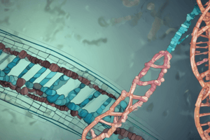
Chromatin
Chromatin
Signup and view all the flashcards
Euchromatin
Euchromatin
Signup and view all the flashcards
Heterochromatin
Heterochromatin
Signup and view all the flashcards
Acetylation
Acetylation
Signup and view all the flashcards
Methylation
Methylation
Signup and view all the flashcards
Barr body
Barr body
Signup and view all the flashcards
Germline mutation
Germline mutation
Signup and view all the flashcards
Somatic mutation
Somatic mutation
Signup and view all the flashcards
RNA polymerase II
RNA polymerase II
Signup and view all the flashcards
TATA box
TATA box
Signup and view all the flashcards
Barrier sequences
Barrier sequences
Signup and view all the flashcards
RNA interference
RNA interference
Signup and view all the flashcards
RT-PCR
RT-PCR
Signup and view all the flashcards
Neoplasm
Neoplasm
Signup and view all the flashcards
Malignant tumor
Malignant tumor
Signup and view all the flashcards
Histogenetic classification
Histogenetic classification
Signup and view all the flashcards
Teratoma
Teratoma
Signup and view all the flashcards
Hamartoma
Hamartoma
Signup and view all the flashcards
Low-grade tumor
Low-grade tumor
Signup and view all the flashcards
Tumor grade
Tumor grade
Signup and view all the flashcards
Ki-67
Ki-67
Signup and view all the flashcards
Carcinogenesis
Carcinogenesis
Signup and view all the flashcards
Tumor grading
Tumor grading
Signup and view all the flashcards
Tumor staging
Tumor staging
Signup and view all the flashcards
Retrovirus genes
Retrovirus genes
Signup and view all the flashcards
Retrovirus advantage
Retrovirus advantage
Signup and view all the flashcards
Tax oncoprotein
Tax oncoprotein
Signup and view all the flashcards
Determining of the existence of nonviral oncogenes
Determining of the existence of nonviral oncogenes
Signup and view all the flashcards
Transfection
Transfection
Signup and view all the flashcards
Driver genes
Driver genes
Signup and view all the flashcards
Growth factors
Growth factors
Signup and view all the flashcards
Reoxygenation
Reoxygenation
Signup and view all the flashcards
Study Notes
Molecular Basis of Cancer
- Alleles are different versions of genes
- An organism is heterozygous if it has two different alleles
- The cell cycle consists of a long interphase, where genes are expressed and chromosomes are replicated
Chromosomes
- Chromosomes remain together as sister chromatids during interphase
- Individual chromosomes are only distinguishable during mitosis
- Sister chromatids condense into mitotic chromosomes during mitosis, which are then separated into two daughter cells
- Each chromosome is an individual unit that replicates, separates, and partitions into daughter cells
- Three types of specialized nucleotide sequences in DNA control these functions
- Centromere DNA sequences allows each copy to be pulled into daughter cells during division
- The kinetochore is protein complex, attaches chromosomes to the mitotic spindle
- Telomeres are specialized DNA sequences forming the ends of chromosomes.
- Writer proteins mark nucleosomes, while reader proteins recognize these marks and bind tightly to nucleosomes
Chromatin
- Is a complex histones and nonhistone chromosomal proteins
- Binds DNA to form chromosomes
- Nucleosomes use ATP to move DNA relative to the histone core
- The histone code involves writers adding modifications, erasers removing modifications, and readers recognizing and recruiting other proteins
- Euchromatin is less condensed, heterochromatin is more condensed
- Chromatin is folded in loops and coils
- Acetylation loosens chromatin to activate transcription
- Methylation activate or repress transcription, depending on its location
- Phosphorylation and ubiquitination activates transcription
- Methylated CpG islands cause gene silencing, while unmethylated CpG islands act as active promoters
- DNA methyltransferase mostly performs this process
Barr Lichaam
- Is an inactive, compacted X chromosome
- Mutations can be inherited through the germ line
- Somatic mutations aren't heritable
- Each centromere pulls one copy of a chromosome to a daughter cell
- A promoter's activity can be studied using whole-genome sequencing or second-generation sequencing
Polymerase II
- Transcribes genes encoding proteins
- Transcription factor TTD binds to the TATA box close to the promoter, which then allows the DNA helicase to unwind the DNA, giving access to the template strand
- After transcription, mRNA undergoes splicing, capping, and 3' polyadenylation
- mRNA exits the nucleus through pores, and then translated into a protein
Regulation of Gene Expression
- Regulation of gene expression dictates when and how often a gene transcribed
- RNA processing, transport, and localization control
- Translational and mRNA degradation control
- Protein activity control
- Transcription regulators regulate transcription of genes
- Coactivators and repressors bind to transcription regulators on DNA to regulate gene transcription
- Barrier sequences prevent the spread of heterochromatin, and insulator DNA regions prevent cis-regulatory regions from inappropriately activating genes
- RNA interference machinery can selectively shut off the synthesis of target RNAs
- The RITS protein complex modifies histones, directing heterochromatin formation, so that transcription is prevented
- piRNAs transcriptionally silence transposon genes and destroy any RNA produced
- Quantitative RT-PCR used to measure individual RNA amounts in cells
- A DNA microarray can be used to analyse which mRNAs are produced in a cell
- mRNA is turned into cDNA and labelled fluorescently, then hybridize to fragments, then a microscope reveals which mRNA was present
Transcription
- Controlled by gene control regions
- Silencers bind repressors
- Insulators form circles around genes and promoters, preventing enhancers from reaching them
Analysis of Genetic Defects
- DNA Cloning: accomplished by inserting a DNA fragment into a purified genome of a self-replicating genetic element
- Purified plasmid DNA circles = cut with restriction nucleases to create linear DNA molecules
- DNA ligase adds foreign DNA to the plasmid
- Recombinant DNA circles are added to bacteria, which grow and replicate the recombinant plasmids with the foreign DNA
- Cells are lysed, and the plasmid DNA is extracted
Gel Electrophoresis
- Separates DNA by size
- DNA is stained for band visualization
Hybridization
- Detects specific nucleotide sequences
- Labeled probes are added and visualized where they bind, indicating the presence of specific gene sequence, FISH
Southern Blot
- Electrophoresis with probe hybridization detects specific DNA sequences
- DNA is separated by size, the membrane is blocked, and a probe is added that binds to specific sequences
Northern Blot
- Gen expression is studied using RNA
- Separates with gel electrophoresis, adds a probe, then bands are analyzed
PCR
- In vitro gene cloning, ssDNA, primers, hybridization, translation with TAQ pol, and DNA amplification, resulting in a band on the blot due to same length DNA fragments located on the membrane since they span the primers
- qPCR quantifies a given sequence in a sample based on labeled color intensity, reflecting gene experience
Sanger Sequencing
- Determines the seq of DNA or RNA
- Occurs by hybridizing a primer to DNA, adding excess dNTPs and DNA polymerase, then adding one type of base into four different tubes, then observing length of fragments which stop due to ddNTPs, then band patterns reveals the sequence
CRISPR/Cas9
- Modifies genome
- Transfects cells with Cas9 protein and guide RNA plus plasmids containing DNA for these proteins, this also replicates the plasmids in which it is contained
- The proteins then cuts the genome in the HR region which causes specific mutations to occur to these genes it contains
Mass Spectometry
- Identifies which amino acids are sumoylated via analyzing ions from a sample which are separated the ratio of mass to charge to view how frequently they all appear
- SUMO means "small ubiquitin-like modification."
SNP Array
- Detects polymorphism within a population as well as deletion and duplications
- SNPs are small polymorphisms that differ by one nucleotide, also identifies loss of heterozygosity
Fusion Analysis
- Analyzes fusions between two separate genes
- Uses fluorescence in situ hybridization for fusions with an area already documented, NGS to sequence for fusions that are new
The Nature of Cancer
- A neoplasm is a tumor, but a tumor is not necessarily neoplastic
- Cancer is a malignant tumour
- Behavioral classification is either benign or malignant
- Histogenetic classification is a cell of origin; morphology like different tissue types. Everything with the suffix -oma is benign. Adenoma follows glands, papilloma finger like projections, a polyp is not a true neoplasm. Benign mesenchymal neoplasms; lipoma, chondroma.
- Everything with -sarcoma is malignant
- Germ Layers: teratoma, hamartoma, malignant or benign
- Disorganized mass of tissue whose cell types are native to the site of the lesion are known are hamartomas
- Misnomers that have -omas but are actually malignant; melanoma, seminoma, lymphoma
- Morphology immunohistochemistry and molecular analysis are how benign vs malignant cells are typed
Grading Tumors
- Low grade tumors have a better outcomes
- The grade of tumor is degree to which it is different or mature as viewed through a microscope, this involves growth patterns, the extent of differentiation, and nuclear aspects and cell cycle information
- Ki-67 gives cell cycle information, as it is present in all proliferating cells
- G1 = well-differentiated (low grade), G2 = moderately differentiated, G3 = poorly differentiated (high grade), and G4 = undifferentiated
- Objective grading; DNA aneuploidy , TP53 mutations (helps preserve genome), and gene expression profile
Tissue Structure
- Normal tissue contains an epithelial cell layer - basement membrane - stroma (supportive connective tissue cells)
- Squamous cell carcinomas arise from epithelial cells that serve as barriers
- Adenocarcinomas arise from epithelial cells that secrete products into vessels they contain
- Mesoderm produces the connective tissues forming and immune cells
- Mesoderm is derived from cells in the system, CNS and PNS
- Melanomas come from neural crest melanocytes
Hyperplastic growths
- Deviate from normal cells, excessive numbers of them
Metaplasia
- Normal cell layer displaced by one of its neighboring cells
Dysplastic
- Appearance of cells no longer standard
- Hyperplasia and metaplasia are flexible and dysplasia causes mutations and invasion
- Genetic mutations and invasion are not reversible
- Key irreversible events: Loss of tumor suppressor genes, activation of oncogenes, chromosomal instability/aneuploidy
Types of Tumors
- Monoclonal tumour is derived from one single ancestor, while polyclonal derives from genetically distinct subpopulations
- Origin is from monoclonal cells
Procarcinogens
- Dramatically different rates of cancer across the world and the world
- Environmental factors, physical factors, and lifestyle all increase the risk of it occurring
- Some of the cancer is also caused by screening, early detection; factors, include chemical factors.
Rous Sarcoma
- Discovered to lead to cancer
- Hen experiment, after cells were infected a petri dish to isolate the species causing the disease, led to the understanding of the nature of cancer and viruses
- Foci formed; clusters of cells in a petri dish
- Viruses cause it to kill their host cells (virulent), while or allowing the cells to not die, but not long-term with limited viability for an intermediate rate of time.
- Normal cells in a petri dish stop because of their inability to connect with other cells
- Genome of RNA cells can be manipulated to do this, and can stop at a time when there is much layering occurring
Viral Transformation
- Transformation phenotype was transferred from initially infected transformed chicken cell to its descendants
- 41degrees reverts cell to the shape and growth patterns from its past experience
- The cell reverts if it doesn't want the disease in the first place
HPV
- Protein coat and caries a DNA genome
- Closely related viruses SV40 and the mouse polyomavirus and Shope´s Papillomavirus: groups of DNA tumor viruses
Cell Culture
- Normal cells need certain factors to live
- Cancer cells can live independently without factors
- They can also live for an unusual amount of extended period of time
- Normal cells need a substrate, called “anchorage dependence” to live
Viral Integration
- DNA virus in a host cell becomes transcribed in genomes for cells that divide
- HPV integrates an oncogene withing the genome to replicate cancer cells.
Retroviruses
-
Convert their RNA back to DNA
-
Three genes are in viral rep
- 2 for the virus to assemble and function
- 1 to transform the cell into a cancer cell
-
Viral V - src is closely related to the c - src in the chicken and it has a drastic function shift
Retroviruses
- Evolved pre-existing cellular DNA for their own use
- MC29 was acquired to have a cellular genome, termed V - myc oncogene
- These have a high tumorigenic potential in infected chickens Myc had its genes remodelled to gain oncogenic characteristics
The Classes of Tumors
- Retroviruses grouped
- 1 that induces cancer rapidly and carry oncogenes SRC for an example -2 carry oncogenes slowly for tumors proto-oncogenes (ALV → myc, MLV),
- 3 T - lymphotropic leukemia virus which gives cancer encoded in the viral genome with no known oncogenes
In order for bacterial cells to cause cancer, specific criteria has to be meet.
- H pyloni, the cell has to allow a protein, CAG a bacteria product to inhibit the gut, therefore reducing cell to cell signaling, to the effect of enhanced proliferation, apoptosis inhibition.
- Testing tumorigenicity with mice via impairing its immune system
Cellular oncogenes
- To Determine if nonviral oncogenes exist chemically transferred, if so they are then transplanted into mice.
- Foci Were created on the body; meaning its potential to cause a cancerous outcome.
- DNA was extracted from those which were 3 - MH, transformed mouse cell lines, into NIH, 3T3 recipient which also formed foci, and were anchorage and tumorigenic
Human Tunor
- Oncogenes which were retro virus associated were present at high number, by amplifying cell genomes. increased amount of potein to to cancer cell
- erbB2: amplified form often increased amount of potein which drives a breast cancer
- Development: the growth of Tunor can cause passengers with causal issues with development
- Genes responsible for the loss of growths
- The normal regulation might enhance
Point mutation for cancer
- By analyzing site structure and looking molecular restriction nucleuses and their site analysis. In the H-ras
- Where one G was swapped out for the proto-oncogene with a T in oncogene
K-ras
- Protooncogene regulatory sequences, controlling for expressions
- Which causes substitution of amino acid
- An normal gene can be turned into a oncogene by the number of copies it expresses, due to the translocation of the proto- oncogene.
- Mhyc gene occurs through translocations, and its also activated
- Translocations is done by deleting miRNA which prevents the cell destruction. can also cause a gene to be ocogenic by deletting sites for mirna
- Translocation create hibrbrid poteins and change their functions
- Growth stem signals can be sent even if EGF receptors are taken away
Growth factor & the pathway
- Gleevec is a selective tyrosine kinase inhibitor which drives proliferation
- Growth factors are proteins sent to give messages to other cells
- These GF’s are also know as mitogens, and this allows an ability to have the cells able to proliferate
- Which leads to dramatic changes in cell expression Src: functions to remove phosphate from 5 ATP and transfers it to a portein substrate, to then be covalently bound with more acid chains The EGF & the pathway
EGF-R
- Protrudes into the extracellular space
- Transmembrane is in the hydrophobic volume which turns the membrane into the plasma
- The ligand stabilizes at some point they are monomers
- Then phospyrolates the tyrosine, leading to the signals
The Jaks polypeptide Cytokine
- Responds through ligand bining
- TGF-B function as Herter Dimmers
- Notch 1 and notch 2 receptors
- Patched, which are primary for the cilium’s
- wnt Factors Ras: can go for both an anchored cytoplasmic membrane and go into the cytoplasm and then to its core.
- Ras activation is triggered by exchange GEF as inactivation is set forth
- Lack of GTPase Activity, a mutation will make it continue even the event its purpose is already fulfilled by it
- Immediate early genes MyC: protein rises after stimulation but decrease as stimluation is gone, due to its short half life, to be replaced by src, the proto is created
- This makes the proto more vulnerable
Protein Activation
- Shc: for ras activations then becomes a bridge between others factors & proteins which then allows a tyrosines to create a phosphate compound of the tails
Grb-2: then activates receptors, which then the SOS allows the RAS to activate downstream Raf: to the serine
Akt molecule
- Become tethered to the PIP3 to reach PH Domain
- Activation creates a stimulus of prolifiting cells with higher chance of metastasis
Cancer Cell proliferation
- Driven by cancer ce dimerizaiton Jaks separate poly peptides
- Jak's phosphorylation activates c receptors
Rora Responds to winths
- B - catenin is the most important , it is what has the most protien loss in the event of cancer.
Tumors
- If cell is absent in the absence of proliferation they will undergo APO - kins
- In a petri dish, cells tether which the body creates and forms the stability for the cells in it
- Ras is able to mimic signals
Cytooplasmic Signaling
- A GEF for Ga, leads stimulates of cell proliferation
- Duals pathways operate then dispatchs protiens to normal cells
Tumor Suppressor Genes
- Cells were in fused whewre alleles were forced to co exisit, synctiyumn of two cells from various origins
- Tumor cells fused with normal cells create a lost to form
- Malignant phenotype was recessive
Point
The cell is to lose its genes. So in that sense they are more prone to the spread than that to start tumor, The tumor can not cause enough if the body can not have its defenses. If first allele is inactivated by chance mutation, the second can be lost during recombinations or the chromosomes themselves.
How the allele is caused the cause
-
Through DNA being elongated during cell division
-
This can cause the disease to come out
-
13q14, for what part in the body of the receptor is responsible
-
Restriction fragment lengths, help show the lOH SNPs with PCR helps complement CpG methylation cause gene silence in these type of cancer mutations They are related to gtp-ase activating
Familial Adenomatour polposis
- Has APC as well Enterocyte encounter
- What they must have for there to be loss
Capable of Induce
- DNA damage regulated signals
- P53 activation on a TF after sythesizing the TF and then destruction
- Survival signals activaie the alkt leading to tumor cell surivival and or growth PI3: gets activated
E2F Activity
-
Leading to AF is an expression
-
P3 leads inhibits the function
-
It is very hard to study because it spreads so commonly and so easily
-
Attrition : a tumor that comes and goes depending on the number of factors at play and to what effect that it is being triggered is more than the cause.
Hemotiximality
- By the flow if you understand you’ll undersetand there importance. to each individual.
Studying That Suits You
Use AI to generate personalized quizzes and flashcards to suit your learning preferences.
Related Documents
Description
Explore the molecular basis of cancer with a focus on chromosomes. The cell cycle includes a long interphase where genes are expressed and chromosomes are replicated. Discover how sister chromatids condense into mitotic chromosomes during mitosis and are separated into daughter cells. Learn about centromeres, kinetochores and telomeres.