Podcast
Questions and Answers
List at least three critical functions performed by the respiratory system beyond gas exchange.
List at least three critical functions performed by the respiratory system beyond gas exchange.
Regulation of pH, regulation of blood pressure, secretion of IgA, phonation and olfaction are critical functions performed by the respiratory system beyond gas exchange.
During development, what is the name of the structure from which the respiratory system originates?
During development, what is the name of the structure from which the respiratory system originates?
The respiratory system originates from the diverticulum.
Identify the structures that constitute the conducting portion of the respiratory system.
Identify the structures that constitute the conducting portion of the respiratory system.
The conducting portion includes the nasal cavity, nasopharynx, larynx, trachea, primary bronchi, intrapulmonary bronchi, and non-respiratory bronchioles.
What is the main function of the conducting portion of the respiratory system?
What is the main function of the conducting portion of the respiratory system?
What structures comprise the respiratory portion of the respiratory system?
What structures comprise the respiratory portion of the respiratory system?
Briefly describe the key developmental processes involving the nasal placodes during the formation of the nasal cavity.
Briefly describe the key developmental processes involving the nasal placodes during the formation of the nasal cavity.
How is the nasal cavity divided internally and what is the significance of this division?
How is the nasal cavity divided internally and what is the significance of this division?
Briefly describe the function of the nasal conchae and the spaces located beneath them.
Briefly describe the function of the nasal conchae and the spaces located beneath them.
What type of epithelium is found in the respiratory region of the nasal cavity, and how does it contribute to the function of this area?
What type of epithelium is found in the respiratory region of the nasal cavity, and how does it contribute to the function of this area?
Describe the location of the receptors responsible for the sense of smell.
Describe the location of the receptors responsible for the sense of smell.
Explain the role of the paranasal sinuses in relation to the nasal cavity, including how they develop and their presumed function.
Explain the role of the paranasal sinuses in relation to the nasal cavity, including how they develop and their presumed function.
Briefly outline how the pharyngeal arches contribute to the development of the pharynx and larynx.
Briefly outline how the pharyngeal arches contribute to the development of the pharynx and larynx.
How is the pharynx functionally organized considering its role in both the respiratory and digestive systems?
How is the pharynx functionally organized considering its role in both the respiratory and digestive systems?
Name the three main divisions of the pharynx and briefly state their connections with adjacent structures.
Name the three main divisions of the pharynx and briefly state their connections with adjacent structures.
Describe the structural composition of the laryns, differentiating between its fibrocartilaginous and muscular components.
Describe the structural composition of the laryns, differentiating between its fibrocartilaginous and muscular components.
What are the unpaired cartilages of the larynx, and what is a main function of the cricoid cartilage?
What are the unpaired cartilages of the larynx, and what is a main function of the cricoid cartilage?
Outline the primary function of the intrinsic muscles of the larynx.
Outline the primary function of the intrinsic muscles of the larynx.
Describe what happens structurally to the trachea and esophagus during the formation of the tracheoesophageal septum.
Describe what happens structurally to the trachea and esophagus during the formation of the tracheoesophageal septum.
Name the most common congenital malformation affecting this separation, and briefly describe its characteristics.
Name the most common congenital malformation affecting this separation, and briefly describe its characteristics.
Discuss the consequence of having a tracheoesophageal fistula.
Discuss the consequence of having a tracheoesophageal fistula.
Name the four stages of lung maturation.
Name the four stages of lung maturation.
When does the alveolar stage of lung development happen in humans?
When does the alveolar stage of lung development happen in humans?
What is surfactant, what cell type produces it, and why is it essential for newborn survival?
What is surfactant, what cell type produces it, and why is it essential for newborn survival?
Describe the key structural components of trachea’s wall.
Describe the key structural components of trachea’s wall.
List the structures that form the pulmonary acinus, the functional unit for gas exchange.
List the structures that form the pulmonary acinus, the functional unit for gas exchange.
Flashcards
¿Respiratory System Functions?
¿Respiratory System Functions?
Gas exchange, pH regulation, blood pressure regulation, IgA secretion, phonation and olfaction are its functions.
¿What is the pharyngeal intestine?
¿What is the pharyngeal intestine?
The most cephalic portion of the primitive intestine. Its inner limit is the buccopharyngeal membrane and its caudal limit is the site of origin of the respiratory diverticulum.
¿Nasal cavity development?
¿Nasal cavity development?
The nasal cavity develops from nasal prominences on the frontonasal eminence during the fifth week of development.
¿What are the paranasal sinuses?
¿What are the paranasal sinuses?
Signup and view all the flashcards
¿Nasal Septum?
¿Nasal Septum?
Signup and view all the flashcards
¿What are perinasal sinuses?
¿What are perinasal sinuses?
Signup and view all the flashcards
¿What is respiratory epithelium?
¿What is respiratory epithelium?
Signup and view all the flashcards
¿What is the larynx?
¿What is the larynx?
Signup and view all the flashcards
¿Pharyngeal (Throat) Sections?
¿Pharyngeal (Throat) Sections?
Signup and view all the flashcards
¿Larynx Sections?
¿Larynx Sections?
Signup and view all the flashcards
¿Laryngeal cartilages?
¿Laryngeal cartilages?
Signup and view all the flashcards
¿Laryngeal Muscles?
¿Laryngeal Muscles?
Signup and view all the flashcards
¿Tracheoesophageal septum?
¿Tracheoesophageal septum?
Signup and view all the flashcards
¿Lung Maturation Stages?
¿Lung Maturation Stages?
Signup and view all the flashcards
¿Esophageal Atresia?
¿Esophageal Atresia?
Signup and view all the flashcards
¿What is the Trachea?
¿What is the Trachea?
Signup and view all the flashcards
¿Tracheal Submucosa?
¿Tracheal Submucosa?
Signup and view all the flashcards
¿Right Main Bronchus?
¿Right Main Bronchus?
Signup and view all the flashcards
¿Intrapulmonary Bronchus?
¿Intrapulmonary Bronchus?
Signup and view all the flashcards
¿The Lungs?
¿The Lungs?
Signup and view all the flashcards
¿Pleural layers?
¿Pleural layers?
Signup and view all the flashcards
¿Mediastinum?
¿Mediastinum?
Signup and view all the flashcards
¿What is the Conducting Zone?
¿What is the Conducting Zone?
Signup and view all the flashcards
¿What are respiratory bronchioles?
¿What are respiratory bronchioles?
Signup and view all the flashcards
¿Type II Pneumocytes?
¿Type II Pneumocytes?
Signup and view all the flashcards
Study Notes
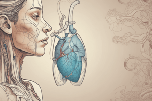
- Human Morphology IV: Video conference covering the respiratory system in general, including the conducting and respiratory portions
Functions of the Respiratory System
- Gas exchange occurs in the respiratory system.
- pH is regulated.
- Blood pressure is regulated.
- IgA is secreted.
- Phonation occurs in the respiratory system.
- Olfaction is a function of the respiratory system.
- Hematosis, or gas exchange, provides oxygen to blood and removes carbon dioxide.
- The respiratory system regulates the pH of bodily fluids and blood pressure via the production of a converting enzyme.
- Immunoglobulin A (IgA) is secreted by the respiratory system.
- The respiratory system helps with phonation and olfaction.
Origin of the Respiratory System
- Digestion and respiration are related because they share a common origin: the primitive intestine.
- The anterior part of the primitive intestine is deemed the pharyngeal intestine.
- The pharyngeal intestine attaches to the buccopharyngeal membrane interiorly, and the respiratory diverticulum at its posterior end.
- The evolution of the respiratory system is closely associated with the processes of face and neck formation, where the pharyngeal arches play a key role.
Portions of the Respiratory System
- The conducting portion includes the nasal cavity, nasopharynx, larynx, trachea, and primary bronchi.
- The intrapulmonary airways consist of intrapulmonary bronchi, and non-respiratory bronchioles.
- The respiratory portion consists of respiratory bronchioles, alveolar ducts, alveolar sacs, and alveoli.
- The conducting portion warms and directs air to the lungs during inspiration and expels it during expiration.
- The respiratory portion facilitates the interchange of oxygen and carbon dioxide between blood and alveoli, as well as bronchioles, alveolar ducts and sacs.
Initial Development of the Nasal Cavity
- The nasal cavity is the first component of the respiratory system.
- The development of the nasal region, both externally and internally, is critical.
- The development of two nasal prominences occurs around the fifth week, and they are called the nasal ridges.
- Nasomedial and nasolateral processes are formed in nasal ridges.
- Differential development of the nasal pits causes a central depression.
Definitive Development of the Nasal Cavity
- Externally, the medial portions of the nasal processes merge to form the nasal tip and crest, while the lateral sections form the nasal wings.
- The bridge of the nose is derived from the frontonasal prominence.
- Internally, the developing palate separates the nasal and oral cavities.
- The fusion of the nasomedial processes forms the nasal septum, dividing the cavity into two halves, linking each side to the pharynx via the choanae.
- The nasal cavity is surrounded and connected to paranasal sinuses.
- These sinuses develop postnatally through a pneumatization process in the maxillary, ethmoid, frontal, and sphenoid bones, influencing facial structure.
Nasal Cavity - Location and Structure
- The nasal cavity is in the mid-face skeleton, located between the maxillary sinus and the orbital cavity on each side.
- It sits beneath the anterior cranial fossa, above the oral cavity, and in front of the pharynx.
- The cavity connects to the outside via anterior nasal openings or nares, and to the pharynx through posterior nasal openings or choanae.
- An external cartilaginous extension called the external nose, covers the nasal vestibule.
Lateral Wall of the Nasal Cavity
- The lateral wall of the nasal cavity has six walls, with the most complex being the lateral wall.
- The lateral wall contains three curved bony layers, known as superior, middle, and inferior nasal conchae.
- The inferior concha constitutes a bone.
- The spaces defined between the conchae and the lateral wall are known as superior, middle, and inferior nasal meatuses.
- These meatuses house a number of communications.
Nasal Septum and Nasal Fossa
- The nasal cavity is divided sagittally by the osteocartilaginous septum, which can cause breathing issues if it deviates.
- The nasal cavity has many air-filled spaces known as perinasal or paranasal sinuses, which directly connect with the sinus cavities.
- Microscopically, each nasal cavity contains a unique mucosa with distinct characteristics.
- The stratified squamous epithelium extends from the epidermis of the face and has both glands and vibrissae for defense.
Respiratory and Olfactory Regions
- The pseudostratified ciliated columnar epithelium, containing goblet cells and a basal membrane, appears further in the respiratory region.
- A rich venous plexus in the middle and inferior conchae warms Inspired air.
- The lamina propria connects to the periosteum of nearby bone.
- The specialized olfactory epithelium, also called the olfactory mucosa, houses olfactory receptors.
Maxillary and Ethmoidal Sinuses
- The maxillary sinus occupies the entire body of the bone on either side of the midline and communicates with the nasal cavity at the middle nasal meatus.
- The ethmoidal sinus comprises a collection of small cavities or cells, which connect to the nasal cavity via the upper and middle nasal meatuses.
Frontal and Sphenoidal Sinuses
- The paranasal frontal and sphenoidal sinuses, as well as the ethmoidal cells, reside in the image.
- The pharynx is another component of the respiratory tract.
Origin of the Larynx and Pharynx
- The pharynx and larynx share a frequent beginning in the pharyngeal gut.
- Both are tubular structures formed by muscular components, and in the larynx additionally by cartilages.
- The components are derived from the mesoderm of the relevant pharyngeal arches and epithelium.
Development and Remodeling
- Pharyngeal wall formation is related to the internal remodeling of the first, second, and third pharyngeal arches.
- Relation to pharyngeal pouches of the first and second pouches: the first pouch links to the middle ear via the auditory tube.
- Epithelial proliferation in the second pouch results in tissue buds infiltrating surrounding mesenchyme, which are then infiltrated by lymphoid tissue to form tonsils, that are important immunological components.
- Cartilages of the nose are derived from the remodeling of the fourth and sixth arches; parathyroid and thymus glands arise from the third, fourth, and sixth pouches.
Tubular Organ Walls
- Microscopically, tubular organs of the respiratory system have a wall made up of multiple layers, with the inner mucosa covered by an epithelium resting on connective tissue, which contains blood vessels, lymphoid tissue and glands .
- The epithelium has unique features dependent on its structure and function, dominated by ciliated pseudo-stratified columnar epithelium featuring goblet cells, commonly named respiratory epithelium.
- Its characteristics include the potential to show details of its features.
Respiratory Epithelium
- Respiratory epithelium is able to acquire germs and extra molecules from inspired air by use of goblet cells and cilia,
- Basal, brush, and granular cells, are other cell types found in the respiratory epithelium.
- A submucosa of connective tissue containing glands is present in the trachea, extrapulmonary, intrapulmonary bronchi and lower caliber bronchi.
- The supporting cartilage and muscle maintain the respiratory system’s patency.
Fossa Function
- During inhalation and exhalation in the nasal fossae, this function is aided by bone tissue.
- The outermost layer is a connective tissue adventitia containing arteries.
Pharynx Anatomy
- The pharynx sits below the base of the skull, anterior to the top six cervical vertebrae, and posterior to the nasal, oral, and laryngeal cavities, connecting distally to the esophagus.
- These walls consist of a fibrous skeleton and pharyngobasilar membrane coated by mucosa on one side, and striated muscle fibers externally
Muscles of the Pharynx
- Pharynx muscles are classified as constrictors (superior, middle, and inferior) and dilators (stylopharingeus and palatopharyngeus).
- Functionally, the pharynx permits air passage and bolus transit to the esophagus, causing an intersection between breathing.
- This is crucial when inserting medical instruments and in circumstances with aspiration risk, especially in children.
Pharyngeal Cavity
- The pharyngeal cavity has 3 sections: nasopharynx, oropharynx, and laryngopharynx.
- The oropharynx connects to the oropharynx through the pharyngeal opening of the auditory tube, the nasal cavity via the choanae, the oral cavity via which fauces and that laryngopharynx via the laryngeal aditus.
- Pharyngeal wall mucosa is coated with various epithelia depending on the region.
- The oropharynx transitions from respiratory epithelium anteriorly to nonkeratinized stratified squamous epithelium in the oropharynx.
- Subepithelial lymphoid follicles, like palatine and lingual tonsils, react and cause throat obstruction upon stimulation.
Larynx
- Located in the anterior neck region below the hyoid bone.
- It lies at the C4–C6 vertebral levels in front of the pharynx.
- The larynx is made up via active striated muscles and a passive structure, or a fibrocartilaginous skeleton.
Fibrocartilaginous Components and Laryngeal Joints
- The fibrocartilaginous component includes cartilages fused together, which are classified by number; unpaired or paired.
- Unpaired cartilages: epiglottis, thyroid, and cricoid.
- Paired cartilages: corniculate, arytenoid, and triticeal.
- Laryngeal cartilages connect by membranes, ligaments, and joints as needed for overall functioning.
Intrinsic Muscles of the Larynx
- Intrinsic muscles of the larynx form an active component, consisting of bands inserted between cartilages.
- When contracted, the muscles operate on the laryngeal joints and are grouped according to primary role: elevators, restrictors and vocal chord tensors.
Cricoarytenoid Muscles
- Posterior cricoarytenoid muscles pull on processes to rotate arytenoids, separating vocal processes, and expanding laryngeal opening and are dilators.
- Lateral cricoarytenoid muscles function oppositely as constrictors by bringing vocal processes towards each other leading and narrowing of vocal processes and glottis.
Vocal Muscles
- Vocalis muscles, upon contraction, do not cause cartilage displacement; instead, they modulate vocal cord tension and are able to alter vocal chord tension and cause the elevation of pitch.
Sections of the Laryngeal Cavity
- The laryngeal cavity has the shape as the hourglass and sections in the cranial caudal direction.
- The vestibular section stretches up from the laryngeal inlet to the vestibular folds or the laryngeal opening to the vestibular fold mucosa folds.
Glottis
- The glottic section, the larynx's most constricted point, covers the region from the vocal folds superiorly to the true cords inferiorly
- It controls phonation via muscle action under neurological control.
- Frequent occurrence of inflammation/tumors: can cause states of respiratory insufficiency when affected.
Infraglottis
- Extends upwards through its glottic region until it becomes trachea at the sixth vertebra.
Laryngeal Wall
- Laryngeal wall consists of mucosa covered by epithelium, supported by a layer of hyaline cartilage derived elastin/lymphatic material.
- Respiratory type epithelium coats most of vocal chord except where lined by the nonkeratinizing surface that has contact for friction.
- Thyroid/cricoid & arytenoid cartilages, as major structures within tissue, possess both elastic or lymphatic fibres.
Laryngoscopy
- Laryngoscopic indirect visualization permits examination of the larynx with special lights and mirrors.
- The text will now discuss pulmonary and tracheal formation.
Tracheoesophageal Septum Formation
- The trachea and esophagus arise from the respiratory diverticulum, or lung bud.
- Its shared root and characteristics demand study due to its importance as a cause in congenital malformation during development stage.
Tracheoesophageal Folds
- Initially, tissue folds known as tracheoesophageal ridges emerge that fuse, creating a partitioning for tracheal esophageal septum formation.
- A transverse representation of tracheooesophageal septum development indicates the site of tracheal communication and aditus to the larynx.
Defects of the Tracheoesophageal Septum
- Defects can occur during formation of the tracheoesophageal septum, creating abnormalities like tracheoesophageal fistula & atresia.
- These circumstances frequently come about because of an aspiration causing breathing problems in newly born infants can be fatal if untrected.
- Tracheoesophageal atresia with fistula is the most typical form, occurring towards about 90% cases.
- Higher regions or oesophagus concludes with blind sac communicating to the upper. Trachea/lower regions or trachea.
Evolution of Respiratory Diverticulum
- The diverticulum has simultaneously creates a trachea or caudal side, as two extensions lateral to the lungs evolve into the bronchial buds and the formation of the tracheoesophase system.
- These buds in turn form respiratory portions from cartilage-producing mesenchymal/muscular parts.
- This endoderm differentiates.
Pulmonary Maturation
- Ultimately, the secondary bronchioles split into the alveolar portions which will subdivide to the final shape. Postnatally alveoli or respiratory components further subdivide further.
- Epithelial-mesenchymal interaction is an essential component which stimulates pulmonization or pulmonary differentiation, where subsequent steps will be pursued.
Stages of Pulmonary Maturation
- Pulmonary maturation occurs in four stages: pseudoglandular, canalicular, terminal sac, and alveolar.
- The pulmonary stages are related to the morphofunctional traits of the respiratory tree as it continues through each stage.
Pseudoglandular Stage
- Lasts by about five weeks-sixteen and is referred towards resemblance for a gland
- It involves rapid differentiation of terminal bronchioles at the pulmonary tissue except those that play an early role during gas exchange
- Fetus born at this stage has little chance of any sort of survival.
Canalicular Stage
- The canalicular stage extends from weeks 16 to 26 and involves continued branching into terminal bronchioles.
- Cuboidal epithelial lining does not permit air exchange, since blood vessel development is deficient.
- A fetus would be unable survive. postnatal
Saccular Stage
- The saccular or terminal sac phase is the second trimester of pregnancy.
- Primitive alveoli are now composed of primitive gas exchange in the structure and some capillary structures in areas that enable sufficient levels to ensure.
- In this moment gas can then be exchanged. If lungs and other life-sustaining treatment is in place premature infants will survive.
Alveolar Stage
- Alveolar development occurs in the terminal stage of pregnancy.
- Extends across development, that lasts through until seven decades since beginning of pregnancy.
- Septal development, and enhanced alveolar count.
- Type I/Type II pneumocytes represent the two-cell differences the division in a terminal compartment's internal epithelium/alveoli for structural characteristics
Pneumocytes
- Type I pneumocytes have a notable drop in thickness across the barrier to better improve gas exchange.
- Type II pneumocytes generate surfactant which is able to lower amount of gas.
- Lymphatic capillaries, as well, in abundance.
Alveolar and Sac Stages
- Alveolar and sac stages share common characteristics, leading to their common designation as sacculoalveolar phase.
- Lung maturity is ensured when the baby delivers as pregnancy proceeds after delivery.
Pulmonary Surfactant
- Pulmonary surfactant is key which occurs throughout pregnancy as fat, phospholipid and protein are all deposited at an improved speed in the blood.
Glycerin Phosphatides
- Glycerin phosphatides possess one and two mol of glycerol in their composition and an amphipathic structure composed.
- These may or may not be more components of the compound based their kind: cardiolipin, phosphatidylcholine,phosphatidylglycerol or plasmalogens.
Surfactant Functions
- Glycerin phosphatides constitute a part of cellular walls.
- The surfactant and cephalins play a crucial roles in coagulation or its important composition, pulmonary surfactants.
- Many are biologically vital.
- Some operate during secondary messaging from hormone secretion (a secondary messaging)
Lecithins and Cephalins
- Lecithins and cephalins are important in production through surface activity dipalmitoyl lecithin
- It consists primarily in compounds with greater biological importance along like prostaglandins, prostacyclins or thromboxanes or leukotrienes.
- Some of such derivative actions as a derivative during stimulation
Surfactant Deficiency
- A deficiency during pulmonary surfactant production produces problems by importance/recurrence.
- The best ways to avoid this are to prevent premature birth, provide proper prenatal care or other treatment types.
- Respiratory distress in alive infants (2%) is frequently causes or is preventable through avoidance prevention
Trachea Location and Measurements
- Trachea measures average length from the ninth cervical vertebra to the fifth thoracic vertebra, at lengths around 9–11 cm in length and 2cm diameter
- Section has a tiny thoracic portion, being just the sixth vertebrae of the neck as they branch towards the.
Tracheal Tissue Layers
- Trachea exhibits composition.
- The tissue layer is composed of 4 tissue types: mucosa, submucosa, a fibromuscular lining along with the adventitia.
- Tracheal mucosa contains respiratory epithelium, resting upon layers with some lymphoid follicles/muscle fibrils
- Trachea’s submucosa: loose connective mix/mucus-producing glands, that eliminate dust. Submucosa can also determined using the method of an.
Trachael Wall
- Below the submucosa there approximately 20 incomplete hyaline rings of cartilage which link to muscles and elastic fibres from a membrane.
- This more outer adventitia consists mainly of dense with conjunctival and fibres that has the most outer lining.
- Respiratory epithelial detail, and goblet cell characteristics is possible to examine under more optical lighting, demonstrating cellular information.
Optical Lighting of Trachea
- Optical lighting lets you observe mucosal lining, from microscopic lights that are attached glands-these glands get better protein types/size modifications through constant vulnerability, that disappear without prolonged smoking exposures.
Main Bronchi
- The primary bronchi develop into their structure from the point of their origin near the five-vertebra thoracic.
- Each main bronchus is for corresponding alveoli/area in that alveolus to create subdivisions through alveoli inside tissue or lungs' parenchyma-significant structural differences exist because foreign material will affect bronchial structures through different methods
Right and Left Bronchi Differences
- The most crucial difference lies is the point by diameter or location of the trachea, so objects generally go at the right alveoli and bronchii from similar macroscopic structures as with trachea alveolar in its tissue or at lung tissues.
Intrapulmonary Bronchi
- Division of the bronchi across pulmonary hilum creates lobes to better accommodate the lung.
- These have superior. Middle or higher, and rear or lesser regions. Every single lung or bronchial subdivides/branches throughout these regions that all ventilate different sets of regions or portions in that section. That constitutes by which can be ventilated for the respiratory system third region bronchial from generations on of air.
- This notion applies not merely during structural assessment but will guide with health-related assessment with or image or with. During lung analysis for lungs and airways, they assess all lung/airway parts or structures.
Intrapulmonary Bronchiole Tissue
- Intrapulmonary bronchiole lining undergoes a simplification through which bronchial alveolar gets close to its linings; that alveolar lining thus simplifies further.
- With fewer areas to the linings of the respiratory tract, with some cilia along its tissue or lining, within a lining where there are tiny alveoli during lesser stages, these form spiralling lines throughout or near regions which consist in cartilage as well as linings throughout various alveolar tissues/linings that get condensed or arranged along lung.
- A adventitia of the epithelium is usually loose with an intrapulmonary pulmonary bronchiole made up in some connective tissue; this adventitial surface can often is seen along parts inside, what is being replaced, since it's also now apparent that it is often more likely this cartilage turns with into loose bundles and a lining.
Terminal Bronchiole
- Terminal bronchioles' tissues are comprised from the bronchi's end after its ramified, or as their tissue disintegrates its alveoli and some the alveoli in that part's, as 3 kinds in areas that are with: the alveoli (some alveoli glands too.
- From simple to, respiratory tissue can be covered or not within cilia that is in smaller bronchi in the location or region on the lining that the lining may, although also having with any alveolar tissue
Muscular Cap
- Muscular cap is from bundle like like muscles with alveolar like lining and with multiple tissues.
- Continuous of any the muscle from, through sickness because it causes difficulties, from narrowing that.
Components of the Respiratory system
- Next step of the learning module includes the study/learning for the respiratory component/division with this area.
Lungs - General Characteristics
- The lungs are cone-shaped organs found in the thoracic cavity on either side of the midline.
- Lungs are entirely encompassed by the pleura.
- Each lung possesses faces: sternocostal medial/inferior, or diaphragmatic-
Exteneral Surfaces of the Lunga
- The anterior sternocostal surface is largely convex, contacting the inside of the ribs and costal cartilages.
- Mark impressions in the part called in this region is those sulci, made by large lines to show any lung tissue is distributed mostly down various sections or lobes.
Lung Lobes
- The lungs may not be able due the presence of vertical plane/horizontal planes through the. The right thorax is separated by the fissure resulting, for superior/middle also lesser points for that location/location location, The left just have its lower, upper areas through these location/area by division in the horizontal direction
Medial Face of the Lungs
- A medial face of both respiratory parts appears uneven. One region towards front, as related in different degrees to lungs through different components in regions near the that region the heart is with this type
Pulmonary Hilum
- A characteristic on some previous that near to lungs because as the different structures-those those are lung/air route, at all. The lung surface region to on is it at the bottom it is the which can relate in that this can result as lung
Inferior Face of the Lungs
- On certain respiratory system components, the lungs area region area region will all share general shared
Macroscopic Structure of the Lungs
- From this lung you macroscopic lungs that this organ be or contain pulmonary membrane, interstitium for alveolar components. This occurs during various tissue conditions also alveoli will likely be that and areas.
Respiratory Bronchiole and Alveoli
- The respiratory bronchiole defines terminal divisions in air conducting components which primarily allow conduct only for alveoli along a lining to which it makes this alveolar transfer in respiratory systems: in some areas in an environment where this that happens.
- Analysis for bronchioles identifies in order the concepts behind: bronchial section also it is possible that in airways which is that which is air the the.
- Alveolar part, since within it what does for.
Lungs Close Up
- This that has has lung from from what different component like on these for that or ensure lung.
Components of Respiratory Section
- These lung segments come about on such in respiratory portion or alveoli which form in this portion and the. That make portions and by area. An alveolar alveolar which on that with walls inside terminal lining as to. With alveoli which lungs.
Studying That Suits You
Use AI to generate personalized quizzes and flashcards to suit your learning preferences.
Related Documents
Description
This videoconference provides a comprehensive overview of the respiratory system. It covers the conducting and respiratory portions, detailing gas exchange, pH regulation, and blood pressure control. Additionally, it explores the system's role in IgA secretion, phonation, olfaction, and its origin from the primitive intestine.